
![]() Contributed by Jeff Kleinberger, MD, PhD and Bryan Rea, MD
Contributed by Jeff Kleinberger, MD, PhD and Bryan Rea, MD
CLINICAL HISTORY
A woman in her late 80s is referred to the hematology/oncology clinic for anemia detected during routine laboratory workup. The patient is asymptomatic for anemia. She has multiple concurrent conditions, including chronic kidney disease, obesity, diabetes, lipidemia, hypertension, hypothyroidism, diverticulosis, and gastric reflux. Her complete blood count shows decreased hemoglobin (8.1 g/dL), decreased hematocrit (24.5%), increased platelets (488,000/uL), and increased red cell distribution width (19.2%) (Table 1). Her medical record shows that her last normal hemoglobin and hematocrit levels were 3 years prior, and the levels have been steadily decreasing over that period. The patient also uses omeprazole chronically for her gastric reflux
Additional laboratory tests are ordered (Table 2). The patient has normal vitamin B12 (730 pg/mL) and iron studies. The reticulocyte count is within normal limits (1.8%). Her erythropoietin level is elevated (86 mIU/mL). Lactate dehydrogenase is normal (215 U/L). Her serum protein electrophoresis studies are unremarkable (data not shown). The patient continues to have no symptoms from her anemia.
Because the laboratory workup is non-diagnostic, a bone marrow biopsy is ordered. Images from the bone marrow biopsy, including select stains, are represented in Figure 1. Flow cytometric immunophenotypic studies performed on the bone marrow specimen are unremarkable (data not shown).
Figure 1: Bone marrow biopsy and aspirate images
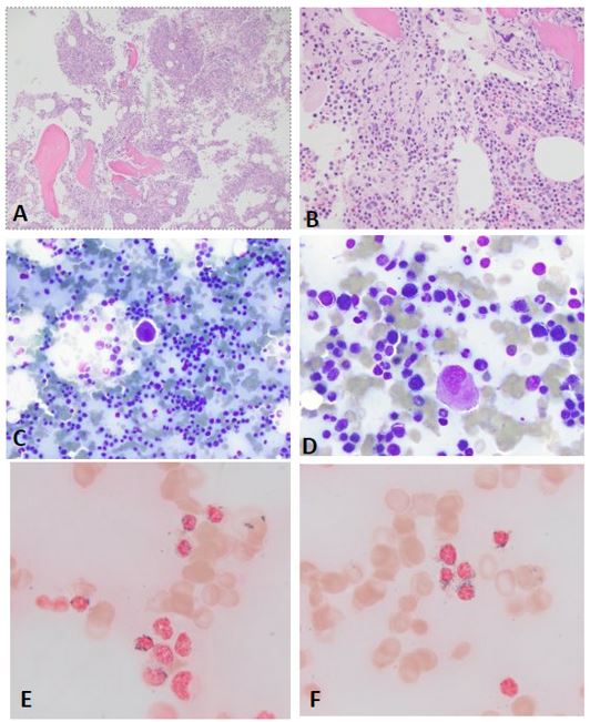
A: Core biopsy (hematoxylin and eosin, 100x), B: Core biopsy (hematoxylin and eosin, 400x), C: Aspirate smear (Diff-Quik, 400x), D: Aspirate smear (Diff-Quik, 1000x), E and F: Aspirate smear (Prussian Blue, 1000x)
What would be your diagnosis based on the laboratory results and microscopic findings? Would any further studies be important?