
IMAGING DESCRIPTION:
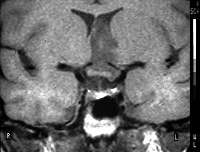
CT and MRI studies revealed a hypointense frontomedial subcallosal mass anterior to the third ventricle. The cortical lesion was relatively well-demarcated. The frontal horn of the left ventricle was slightly compressed; otherwise, no mass effect was seen. The basal parts of the tumor enhanced focally, scantly with contrast medium. Calcification was not observed. Review of previous imaging studies from 1990 until 1995 showed that the appearance of the tumor had not changed.
A stereotactic biopsy was performed followed by subtotal resection half a year later.