
MICROSCOPIC DESCRIPTION:
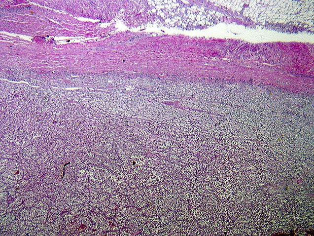
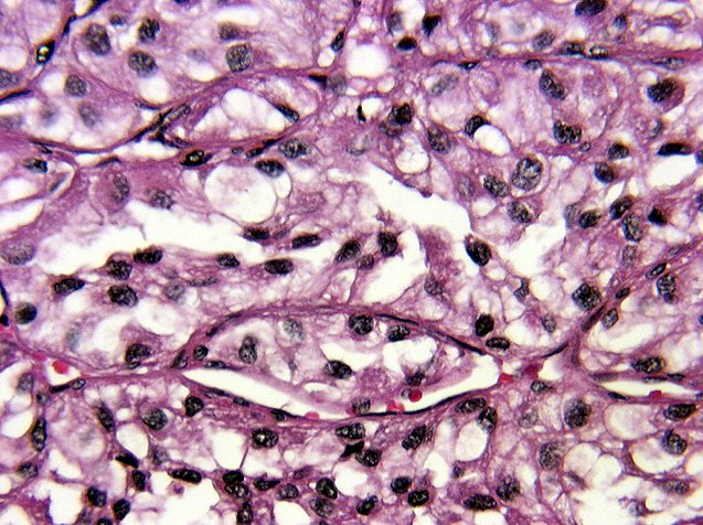
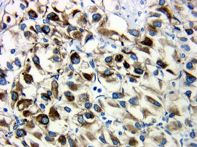
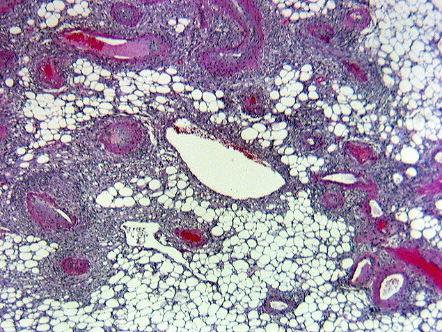
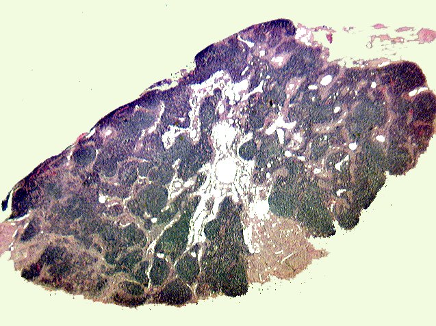
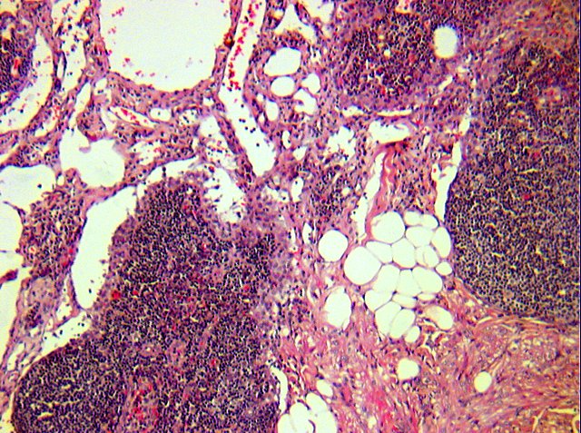
The large tumor mass contained extensive areas of central necrosis and hemorrhage surrounded by nests and trabeculae of neoplastic cells supported by a delicate fibrovascular stroma which contained a minimal amount of chronic inflammatory cells. The majority of the neoplastic cells contain clear cytoplasm although focal areas cells containing granular eosinophilic cytoplasm are identified. The nuclei are pleomorphic with an irregular nuclear contour containing occasional small, single nucleoli and rare mitotic figures. No areas of sarcomatoid differentiation were identified. The neoplastic cells showed strong focal reactivity with antibody against cytokeratins AE1 and AE3 but were uniformly negative when stained with HMB45 antibody. In the adjacent renal parenchyma, focal nodules containing mature adipose tissue, thick-walled blood vessels lacking an elastic lamina and irregular sheets of HMB45 positive smooth muscle found primarily in a perivascular distribution were identified. Similar appearing nodules were present within the perinephric lymph nodes (Image 01 and Images 02).