
![]() Contributed by Waseem Anani, MD; Charleen Chu, MD, PhD; Tim Oury, MD, PhD
Contributed by Waseem Anani, MD; Charleen Chu, MD, PhD; Tim Oury, MD, PhD
PATIENT HISTORY
The patient is a 30-year-old male with a 2-month history of a right corneal abrasion when removing his contact lens. Initial bacterial and fungal culture results were negative, and when he returned for a follow-up appointment, he complained of increasing ocular pain, headaches, and vision loss. Physical exam revealed an abrasion that had failed to heal, leading to a purulent corneal ulcer. Ocular fluid was collected, and the patient was started on antibiotics for a suspected bacterial infection.
The patient then traveled to Florida where his symptoms continued to worsen. While traveling, he was evaluated by a physician, and Polyhexamethylene biguanide, Chlorhexidine, and Valacyclovir were added to include Acanthamoeba and Herpes coverage. When the patient returned home, his symptoms failed to resolve, and he returned for an ophthalmology follow-up appointment. An eyewash specimen was sent for bacterial, fungal, and protozoan cultures as well as Acanthamoeba molecular testing.
The patient's vision continued deteriorating which signaled resistance to therapy. He was taken to the operating room for a corneal transplant. H&E images from his cornea are shown below.
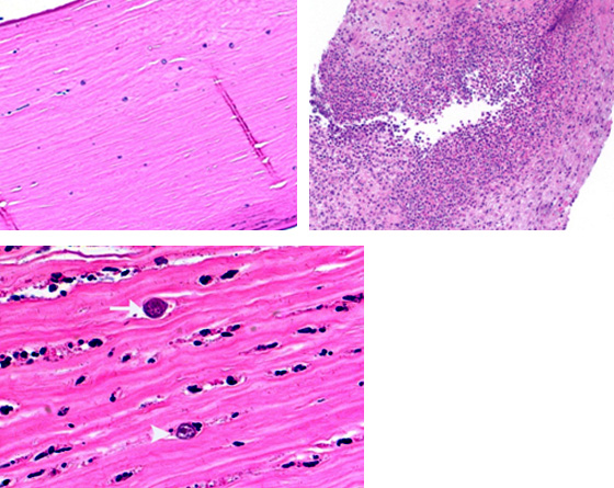
The corneal specimen demonstrated numerous scattered forms (top left) on a background of acute necrotizing keratitis (top right). Many viable organisms were observed superficially and deep with only occasional necrotic forms (bottom left). Two different forms of the same organism were identified: cyst (arrow) and trophozoite (arrowhead).