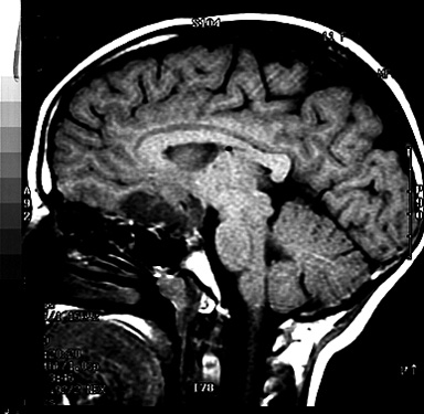
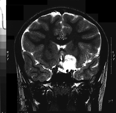
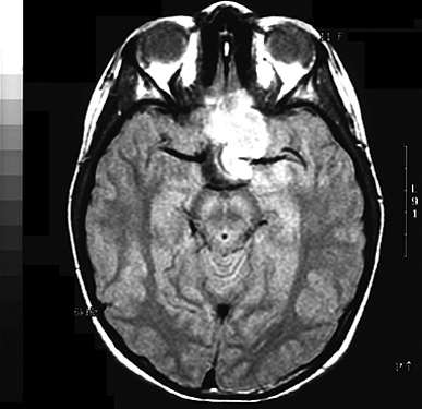
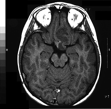
MRI scans demonstrated a tumor in the frontal lobe which was isodense to brain on T-1 weighted images. The lesion was hyperintense in both T-2 weighted and proton density weighted images. After contrast administration, there was little enhancement of the tumor.