

MICROSCOPIC DESCRIPTION:
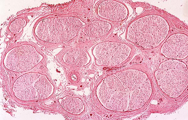
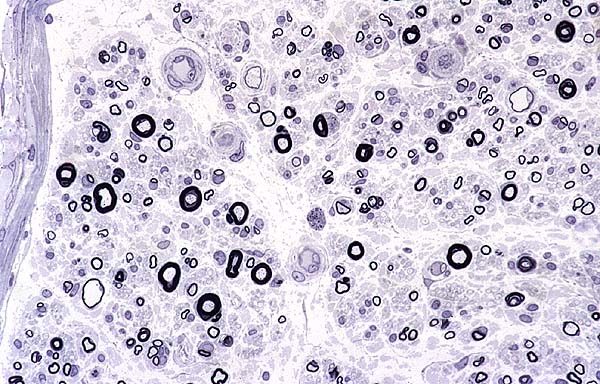
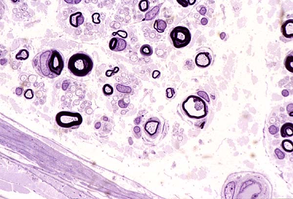
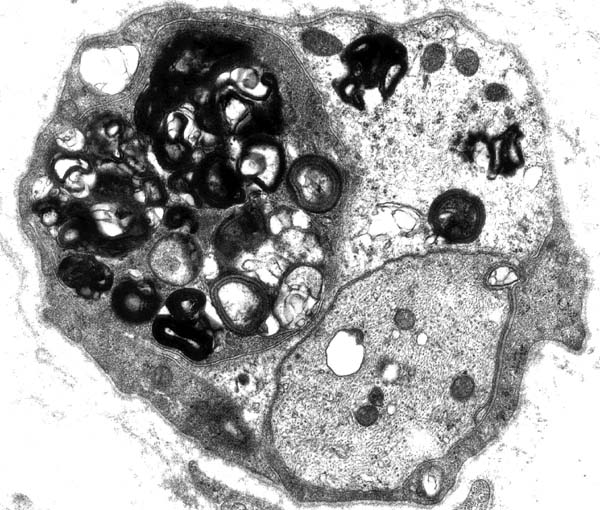
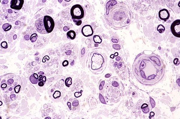
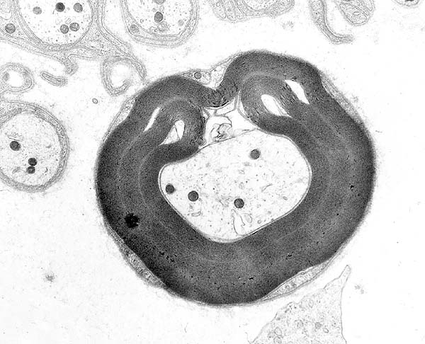
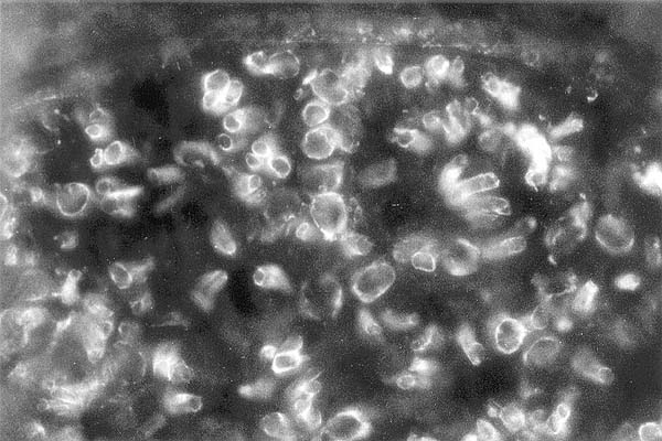
The sural nerve was apportioned for paraffin embedding, "quench" freezing in liquid N and plastic resin embedding. The microscopic anatomy is preserved with 14 fascicles identified (Fig 2). There is no inflammation and congo-red stain shows no amyloid. The fiber density is decreased associated with axonal degeneration (Fig 3), axonal atrophy (Fig 4), segmental demyelination (Fig 5), onion bulbs (Fig 6) and tomaculae(Fig 7). Immunostaining on fresh frozen tissue discloses intense positivity (Fig 8) and transmission electron microscopy shows widespread abnormality of myelin periodicity (Fig 9 and Fig 10).