MICROSCOPIC DESCRIPTION:
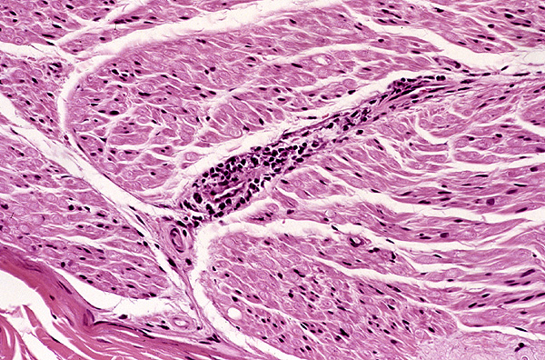
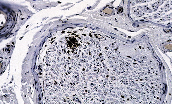
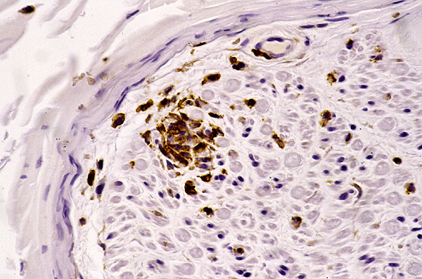
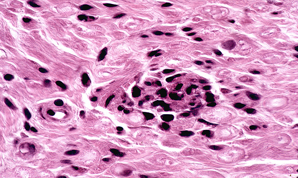
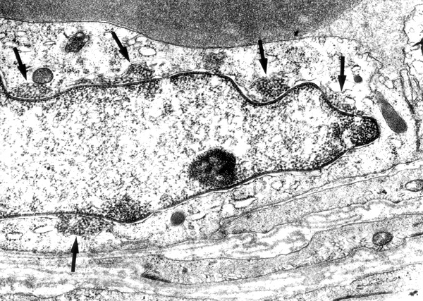
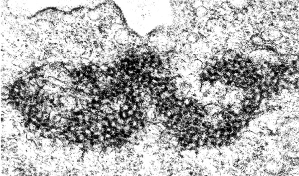
Nerve biopsy showed discrete sleeves of lymphocytes around epineurial and perineurial vessels (Fig. 1) and dense mononuclear cell aggregates obliterating endoneurial microvessels (Fig. 2 [LCA immunostain], Fig. 3 [LCA immunostain] and Fig. 4 [H&E, oil immersion]) associated with slight axonal degeneration. Vessels larger than 60 microns were unaffected. When viewed under the electron microscope, perinuclear skeins of tubules were plentiful in endoneurial vascular endothelium (Fig. 5, arrows, note Weibel-Palade body; x19,200 ); with 50% of vessels having the abnormality. Muscle biopsy revealed neurogenic atrophy, focal perivascular lymphocytic cuffs, atrophy of perimysial nerve twigs, siderosis in endomysium and inclusions in endothelium (Fig. 6, x 44,800).