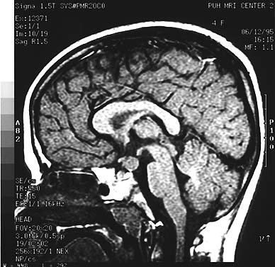
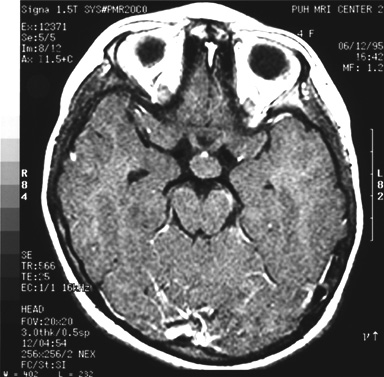
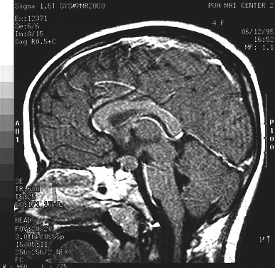
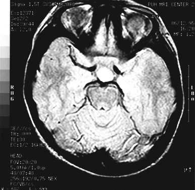
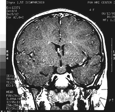
T-1 weighted MRI scans revealed a mass in the tuber cinereum which did not involve the optic nerves (MRI image-1, MRI image-2, MRI image-3). On T-2 weighted images there was a slightly brighter signal than surrounding brain (MRI image-4). There was no contrast enhancement (MRI image-5).