Brain Pathology Case of the Month - January 2018
 Contributed by Alexandros Iliadis1, Elsa Pazarli2, Charalampos-Chrysovalantis Chytoudis-Peroudis3, Stella Chondromatidou4, Ioannis Tsitouridis4, Ioannis Efstratiou2, Dimitrios Kanakis5
1Pathology Department, Medical School, Aristotle University, Thessaloniki, Greece
2Pathology Department, "Papageorgiou" General Hospital, Thessaloniki, Greece
3Department of Molecular Biology and Genetics, Health Sciences School, Democritus University of Thrace, Alexandroupolis, Greece
4Radiology Department, "Papageorgiou" General Hospital, Thessaloniki, Greece
5University of Nicosia Medical School, Nicosia, Cyprus
CLINICAL HISTORY AND IMAGING


This 74-year old woman was admitted to the ER and subsequently our department due to craniocerebral traumata following involvement in a road accident. Brain computed tomography showed right parieto-occipital fracture, left parietal and right occipital subdural hematomas, subarachnoid hemorrhage and diffuse cerebral oedema. Brain magnetic resonance T2 weighted imaging revealed a left parietal, space-occupying lesion (Fig. 1a) with contrast enhancement in T1 weighted imaging (Figs. 1b, 1c). Brain magnetic resonance angiography offered normal findings. In view of the radiologic findings with the identification of the space-occupying mass, the patient underwent excisional surgery to confirm the nature of the lesion.
GROSS AND MICROSCOPIC PATHOLOGY
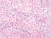
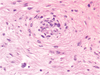
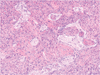
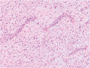
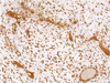
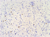
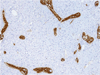
The resected tumor measured 2.8x1.5x1.2 cm and was fairly well circumscribed, whitish in color and fibro-elastic in consistency. Light microscopy showed alternating areas of various stromal consistency and variable cellularity, moderate at most (Fig. 1d). At high power, the mildly atypical tumor cells were fusiform or spindle-shaped, arranged in fascicles, vague whorls or swirls (Fig. 1e), with pale eosinophilic cytoplasm and with mostly vesicular ovoid or angulated nuclei and distinct nucleoli, showing mild nuclear polymorphism and scarce mitotic figures. There were also isolated multinuclear cells as well as lymphoplasmacytic perivascular infiltrations to be seen. The brain parenchyma was regionally infiltrated by neoplastic tongues, while neuroglial islets were present within the tumor (Fig. 1f). Areas with curvilinear vascular network could also be discerned (Fig. 1g) as well as scant foci of necrosis and phagocytosis of the adjacent brain tissue. IHC revealed the following immunophenotype: Vimentin+ (Fig. 1h), EMA--/+ (Fig. 1i), GFAP- (only the above described neuroglial islets were strong positive for this marker; [Fig. 1j]), CD34-, S100-, CK-, SMA- and Desmin-. The MIB1/Ki67 labeling index showed a variable immune-positivity which ranged from 5% in the areas with low cellularity and reached values even up to 30% in the more cellular regions. What is your diagnosis?
FINAL DIAGNOSIS
)
)
)
)
)
)
)
)
)
)