MICROSCOPIC DESCRIPTION:
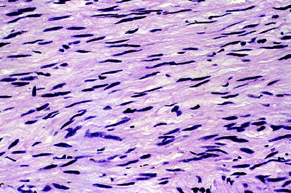
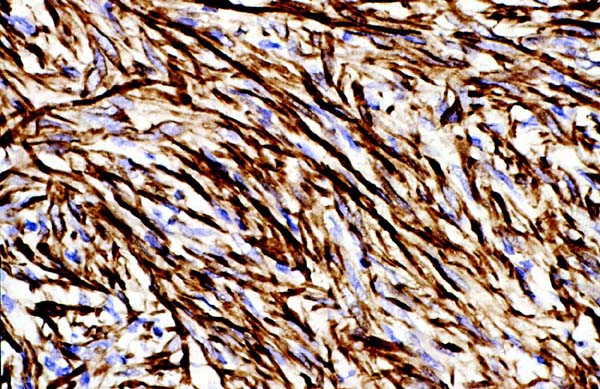
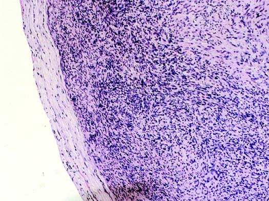
H&E stained paraffin-embedded sections of the intracranial mass showed a moderately cellular spindle cell neoplasm composed of elongated cells with long, slender nuclei containing delicate, evenly distributed chromatin and eosinophilic cytoplasm (Figure 3). Occasional mitotic figures were present. Immunohistochemical stains for EMA and S100 were negative. Similar staining for actin was positive (Figure 4). In-situ hybridization for the Epstein Barr virus genome was also positive. Paraffin embedded sections of the cervical mass showed a similar, but more cellular, spindle cell neoplasm (Figure 5) with a similar immunohistochemical profile and positive staining for Epstein Barr virus.