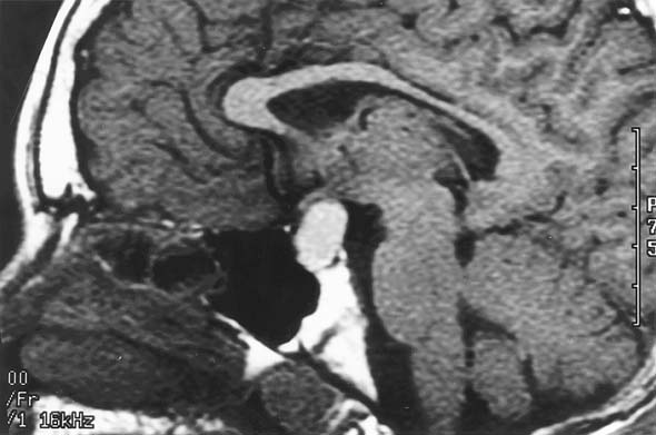

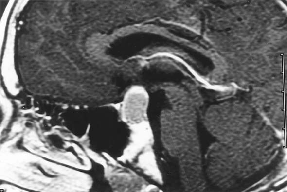
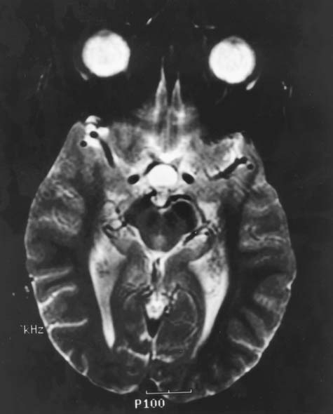
IMAGING STUDIES
Magnetic resonance images (MRI) of the brain revealed an intrasellar and suprasellar cystic mass with well defined smooth borders, which displaced the optic chiasm superiorly. The lesion displayed high signal intensity on T1 weighted images (Images 1a and 1b, sagittal and axial, respectively) and showed peripheral enhancement with Gadolinium administration (Image 1c). The lesion also showed high signal intensity on T2 weighted images (Image 1d). A previous brain MRI from 1991 did not reveal sellar or suprasellar lesions