MICROSCOPIC DESCRIPTIONS:
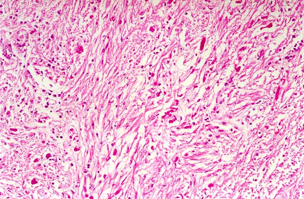
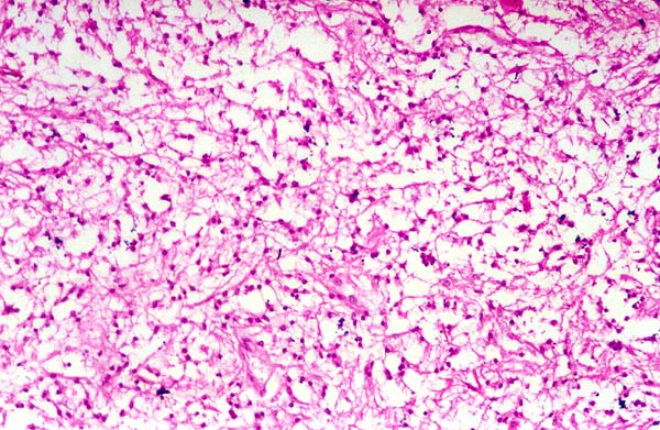
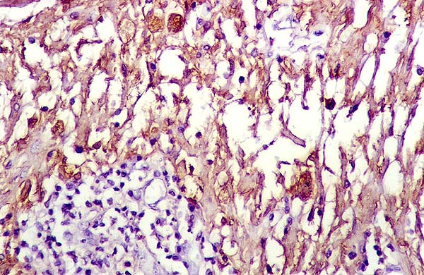
Multiple sections were examined and showed a biphasic tumor consisting of compact areas of elongated and highly fibrillated cells with numerous Rosenthal fibres (Fig.4a) alternating with areas which were composed of loosely arranged cells with microcystic change (Fig.4b). In addition, there were large areas of fresh hemorrhage as well as evidence of old hemorrhage in the form of hemosiderin deposition. Focal lymphocytic infiltrate was also noted. Vessels were thickened and hyalinized but endothelial proliferation was not seen. No necrosis or mitosis was identified. Immunohistochemistry revealed both types of cell to be positive for GFAP (Fig.5).