

MICROSCOPIC DESCARIPTION
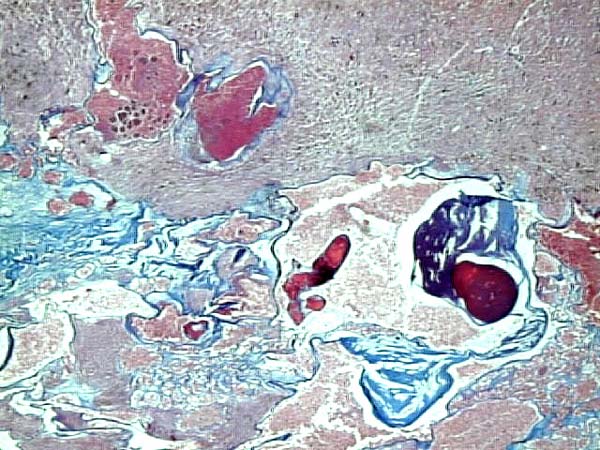
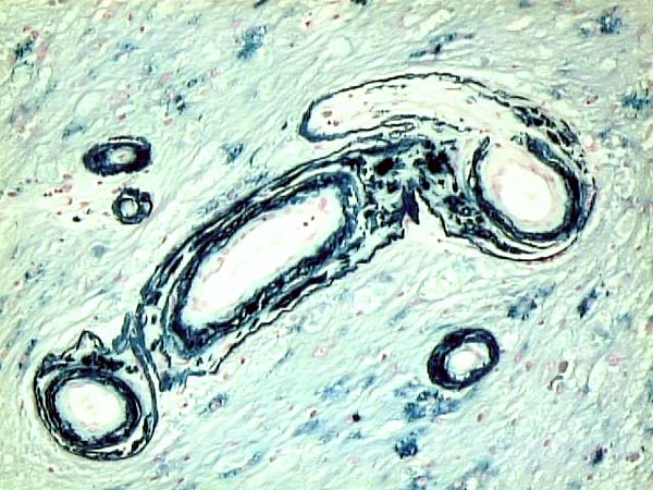
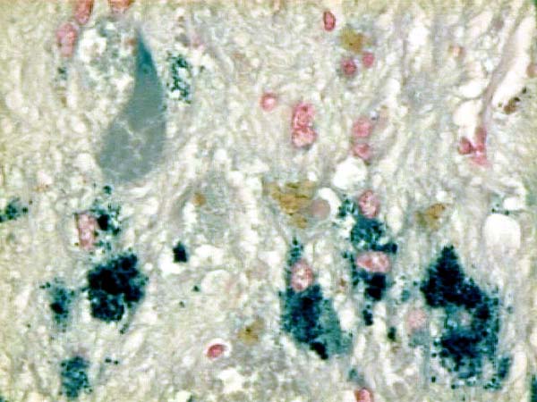
Histologic examination showed numerous vascular channels embedded subcortically in the white matter of the left frontal lobe, clearly seen in this trichrome stain. Venous, cavernous channels were preferentially located in the center of the lesion. The periphery contained numerous arterial branches that were extensively incrustated with hemosiderin as seen in the special stains for iron. (Image A and B). The white matter and cerebral cortex was architecturally distorted, gliotic and extensively siderotic. As demonstrated by the stains for iron, Hemosiderin was predominately in macrophages and astrocytes and, to a lesser extent in cortical neurons as seen in this iron stain.