

MICROSCOPIC DESCRIPTION:
A subtotal resection of the tumor was performed. Multiple fragments of a grayish, partially hemorrhagic tissue were received. Small amounts of tissue were saved for molecular studies and the remainder was fixed in 10% formalin, embedded in paraffin and stained with hematoxylin-eosin (H&E).
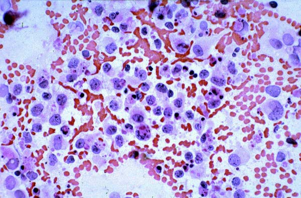
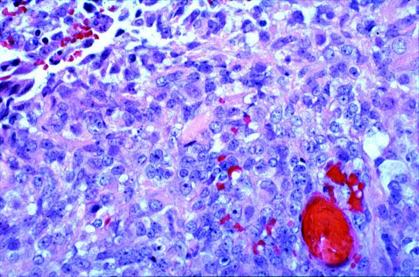
Microscopic examination revealed a cellular neoplasm with multiple foci of geographic necrosis. The majority of the cells had large, vesicular nuclei with open chromatin and prominent nucleoli and variable amounts of cytoplasm, ranging from abundant to scarce. Some cells showed the classic rhabdoid appearance with globular cytoplasm and peripheral nuclei. Mitotic figures were common. There were small areas of primitive appearing cells with high nuclear-to-cytoplasmic ratio arranged in sheets. Typical cytologic and histologic features are seen in these H&E images. The majority of the cells seen in the touch prep are rhabdoid, with a peripheral round to oval nucleus, some of them with prominent nucleolus. Most cells have abundant pink cytoplasm. Areas similar to that represented in the second micrograph (micro.2) composed the majority of the tumor which consisted of rhabdoid cells in sheets, without any architectural organization. (400 x original magnification).
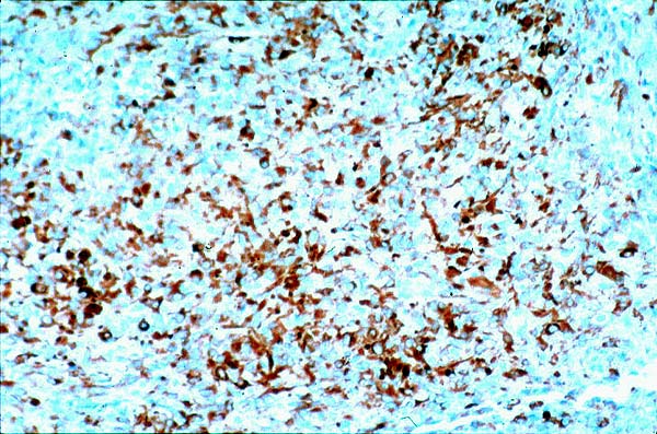
A special stain for reticulin demonstrated abundant staining around individual cells as well as bundles of reticulin dispersed throughout the tumor. A panel of immunostains was performed using standard biotin-streptavidin or ABC methods, and the results are shown in Table I. Representative images of immunostains against vimentin, epithelial membrane antigen and GFAP are presented here. A large proportion of the rhabdoid cells immunoreact against the specified antibodies (200 x original magnification).
| Immunostains | Source | Results |
|---|---|---|
| GFAP | Dako | Diffuse, strong positivity |
| EMA | Dako | Diffuse, strong positivity |
| Vimentin | Dako | Diffuse, strong positivity |
| S100 | Inovation Found | Multifocal, strong positivity |
| PGP9.5 | Ultracin | Multifocal, weak positivity |
| Synaptophysin | Biogen | Focal positivity |
| Muscle Specific Actin | Dako | Focal positivity |
| Desmin | Dako | Negative |
| Neurofilament(three MW) | Dako | Negative |
| CEA | Biogen | Negative |
| Transthyretin (Prealbumin) | Dako | Negative |
| Ki67 (proliferation index) | Dako | 24% |