


MICROSCOPIC DESCRIPTION:
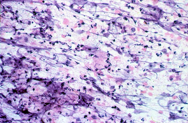
Cytologic examination of direct smears of fine needle aspiration material obtained from the left upper lobe mass revealed a slightly mucoid background, moderate cellularity, polymorphonuclear leukocytes, lymphocytes, and epithelioid histiocytes (image 1 and image 2). Also, a significant number of spherical to ovoid yeast forms were seen which appeared to have a capsule. Several of the organisms exhibited narrow-based budding. Mucicarmine stain highlighted the capsular material. Insufficient material remained to perform Grocott-methenamine silver or acid fast stains.