


MICROSCOPIC DESCRIPTION:
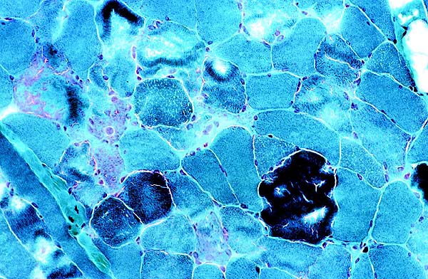
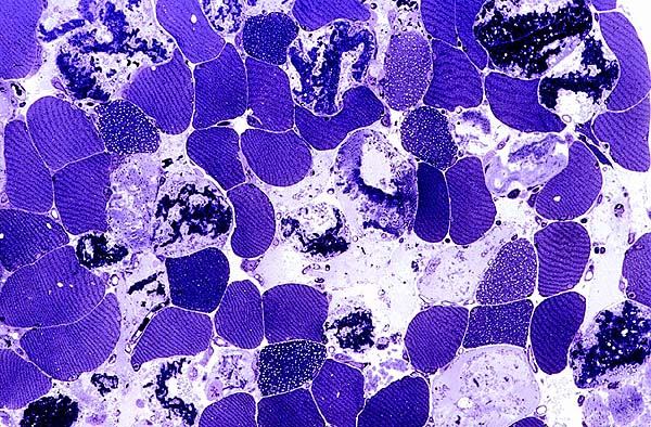
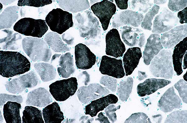
Cryosections stained with hematoxylin, phloxine and saffron (Fig. 1), gomori-trichrome (Fig. 2), and semi-thin plastic resin sections (Fig. 3) showed an abnormality affecting about 40% of myofibers. Early phagocytosis was present in many areas (Fig. 4). There was no inflammation or vacuolar change or perifascicular atrophy. Unaffected fibers had a normal pattern with PAS stain and with myophosphorylase reaction. Both fiber types were affected as shown by ATPase at pH 9.4 (Fig. 5). Immunostaining for leukocyte common antigen revealed no inflammation. Ultrastructural examination disclosed no tubular reticular inclusions (Fig. 6).