
MICROSCOPIC DESCRIPTION:
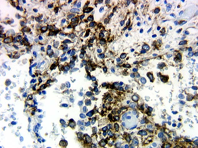
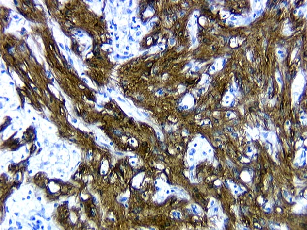
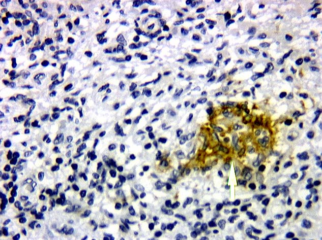
Lungs: Diffuse honeycombing fibrosis was present along with foci of bronchial associated lymphoid tissue with aggregates of hyperplastic histiocytes mixed with the lymphocytes. The histiocytic nuclei were elongated and oval, with irregular membranes occasionally showing grooves or folds (Image 06). These histiocytic aggregates demonstrate positive immunoperoxidase staining for CD1a (Image 07), and S100, and negative staining for CD68.
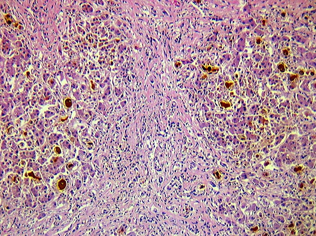
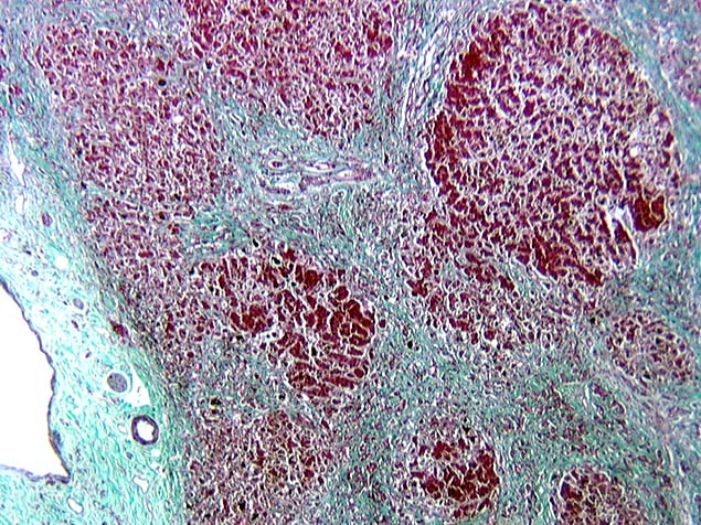
Liver: Extensive micronodular and macronodular cirrhosis was present with bile duct proliferation, bile plugging, and ongoing necrosis of hepatocytes at the periphery of regenerative nodules (Image 08, and Image 09 - trichrome stain). As seen in the lung, focal lymphoid and histiocytic aggregates were identified in the fibrous bands; however, these cells were nagative on CD1a immunostaining.
Brain: The optic chiasmatic mass involving the adjacent hypothalamus showed extensive astroglial proliferation with the astrocytes arranged in "tight" fascicles. The astrocytes had elongated, pleomorphic nuclei and a moderate amount of fibrillary wavy cytoplasm. Associated with the astrocytic mass were a few foci of mononuclear cells and histiocytes in a perivascular distribution (Image 10). The extensive reactive gliosis was demonstrated by a GFAP stain (Image 11). Similar astrocytic proliferations were seen in the pineal body and area postrema of the medulla with prominent Rosenthal fiber formation. Small clusters of the histiocytes again demonstrated positive immunostaining for CD1a (Image 12).
ULTRASTRUCTURAL STUDIES:
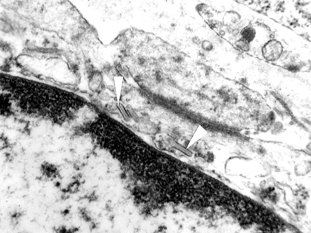
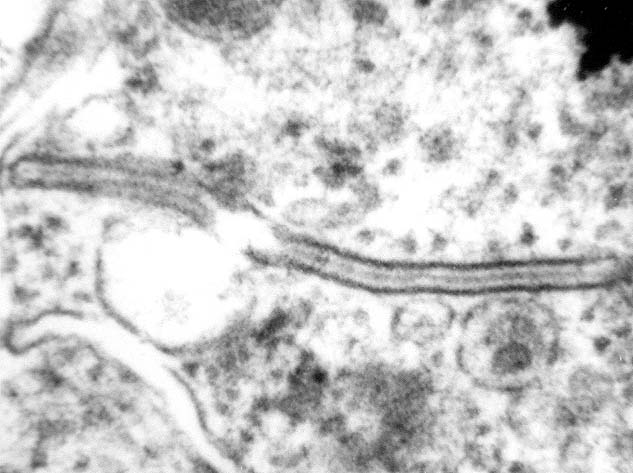
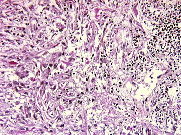
Electron microscopy was performed on sections of the formalin-fixed lung containing to the histiocytic aggregates which immunostained for CD1a. Birbeck granules (believed to be invaginations of the cell membrane) were identified. These granules were usually located near the Golgi apparatus of the cell, appearing as rigid tubular structures with an average diameter of 34 nm, and a zipper-like central core (Images 13 and 14).