![]() Contributed by Pranav P. Patwardhan, MD, and Gabriela M. Quiroga-Garza, MD
Contributed by Pranav P. Patwardhan, MD, and Gabriela M. Quiroga-Garza, MD
CASE PRESENTATION
A man in his sixties presented to the hospital with history of urothelial carcinoma. The patient was status post radical cystoprostatectomy with neobladder creation 8 years prior to the current presentation. Subsequently, the patient had multiple recurrences of the carcinoma involving the ureter which was surgically managed. During the present presentation, the patient had intermittent hematuria and was also found to have a penile lesion. Both urethral and penile lesion biopsies were performed and diagnosed as urothelial carcinoma and extramammary Paget disease, respectively. A partial penectomy was the recommended treatment option and was subsequently performed. The penectomy specimen was evaluated for the presence of the disease and any other pathologic findings.
HISTOPATHOLOGIC EVALUATION
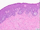
A partial penectomy specimen consisting of glans penis with attached urethrectomy was received. A white to yellow, rubbery, ill-defined and infiltrating, vaguely nodular mass (7.4 x 1.2 x 0.5 cm) was identified surrounding the urethra in the proximal, mid and the distal one-third. The glans penis, the urethra and the corpus spongiosum was grossly involved by the mass while the minimal corpus cavernosa seen for evaluation was not involved. Microscopic evaluation confirmed the presence of recurrent, invasive papillary urothelial carcinoma, high grade arising from a background of multifocal urothelial carcinoma in situ. The carcinoma extended throughout the length of the penile urethra and invaded into the adjacent corpus spongiosum, adipose tissue and glans. No definitive lymphovascular invasion or perineural invasion was seen. Also noted was extensive Paget disease on the glans surface (Fig. 1). On immunohistochemistry, the Paget cells were positive for CAM5.2, CK7, p63, GATA3, uroplakin II/III, while negative for CK20, monoclonal CEA, GCDFP-15, S100 and Melan A. (see Fig 2-6)

Figure 1. H-E Image showing Paget cells (click each image to enlarge)