
![]() Contributed by Azfar Neyaz, MD, and Ivy John, MD
Contributed by Azfar Neyaz, MD, and Ivy John, MD
CASE PRESENTATION
A female in her 40s with no significant medical history presented with a slowly growing, long-standing soft tissue mass in the left lower thigh.
RADIOLOGIC FINDINGS
Magnetic resonance imaging revealed four masses, ranging from 0.4 up to 1.7 cm, involving the rectus femoris and vastus lateralis muscle. The masses were well-defined with isointense T1 and slightly heterogenous hyperintense T2 signal. Concurrent computed tomography of the abdomen and chest demonstrated multiple lesions in the lungs, liver, and spleen, compatible with metastatic disease.
HISTOLOGIC EXAMINATION
The biopsy and subsequent excision of the thigh mass demonstrated epithelioid cells arranged in a nested and pseudoalveolar pattern (Figure 1A). Vascular spaces were noted focally (Figure 1B). On high power, the tumor was composed of nests of large epithelioid cells with abundant glassy to vacuolated cytoplasm and well-demarcated cell borders separated by fibrous septae (Figure 1C). Notably, the tumor cells were positive for CD31, ERG, and TFE3, while negative for pan-keratin, FOSB, and CAMTA1.
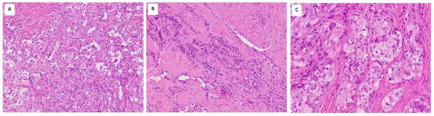
Figure 1. The tumor showed a nested and pseudoalveolar growth pattern (A). Vascular spaces were noted focally (B). The nests of tumor cells were composed of large epithelioid cells with abundant glassy to vacuolated cytoplasm (C).
MOLECULAR STUDIES
Next-generation RNA sequencing analysis (Foundation Medicine, Cambridge, MA, USA) performed on formalin-fixed paraffin-embedded biopsy tissue identifies YAP1::TFE3 fusion.