Final Diagnosis --Clear Cell Papillary Renal Cell Carcinoma (ccp-RCC)
FINAL DIAGNOSIS
Clear Cell Papillary Renal Cell Carcinoma (ccp-RCC)
DISCUSSION
The diagnostic entity of ccp-RCC was described by Tickoo et al. through the analysis of end-stage renal disease associated neoplasms and designated by Aydin et al. in a large series of tumors as clear cell tubulopapillary renal cell carcinoma [1,2]. Diagnostic criteria and terms were integrated in the International Society of Urological Pathology (ISUP) Vancouver Classification of renal neoplasia as ccp-RCC [3]. The entity has been reported to account for approximately 4.3% of all renal cell tumors, the majority of which occurred in kidneys with end-stage renal disease [4]. Although historically associated, the majority of cases are from patients without end-stage renal disease.
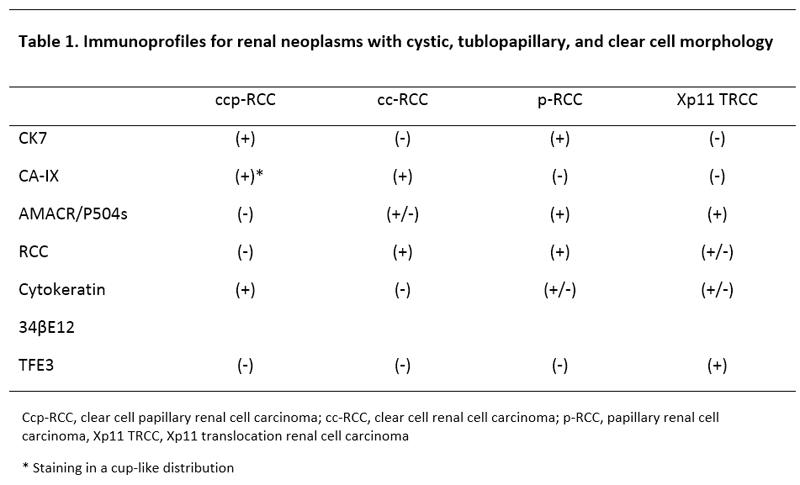
Characteristic histomorphologic findings include a well-defined capsule composed of a mixture of cystic, papillary, tubular, acinar, and solid components. The cells are composed almost entirely of cuboidal or columnar cells with clear cytoplasm and low-grade nuclei arranged away from the basal aspect of the cell (Figure 2). The stromal component consists predominately of fibrous tissue. Tumor necrosis, lymphovascular invasion, and perirenal invasion are absent [4-8]. The immunohistochemical profile of cpp-RCC shows CK7, carbonic anhydrase IX (CA IX), Pax2, Pax8, cytokeratin 34βE12, and vimentin positivity. CA IX staining is in a cup-like fashion (Figure 3C-D). RCC, CD10, AMACR/P504s, parvalbumin, TFE3, and TFEB show negative staining.4-8 Molecular profiles of cpp-RCC have been shown to be distinct from clear cell renal cell carcinoma (cc-RCC) and papillary renal cell carcinoma [8-9].
The differential diagnosis includes kidney neoplasms with cystic, tubulopapillary growth and clear cell morphology including cc-RCC with clear cell papillary-like areas, papillary renal cell carcinoma with clear cell changes, and MiT/TFE family translocation renal cell carcinomas (Xp11.2). An immunohistochemical stain panel can be used to differentiate the entities (Table 1) [8].

REFERENCES
- Tickoo SK, dePeralta-Venturina MN, Harik LR, Worcester HD, Salama ME, Young AN, Moch H, Amin MB. Spectrum of epithelial neoplasms in end-stage renal disease: an experience from 66 tumor-bearing kidneys with emphasis on histologic patterns distinct from those in sporadic adult renal neoplasia. American Journal of Surgical Pathology. 2006;30:141-53.
- Aydin H, Chen L, Cheng L, Vaziri S, He H, Ganapathi R, Delahunt B, Magi-Galluzzi C, Zhou M. Clear cell tubulopapillary renal cell carcinoma: A study of 36 distinctive low-grade epithelial tumors of the kidney. Am J Surg Pathol. 2010;34:1608-1621.
- Srigley JR, Delahunt B, Eble JN, et al. The International Society of Urological Pathology (ISUP) Vancouver Classification of Renal Neoplasia. Am J Surg Pathol. 2013;37(10):1469-1489. doi:10.1097/PAS.0b013e318299f2d1.
- Alexiev BA, Drachenberg CB. Clear cell papillary renal cell carcinoma: Incidence, morphological features, immunohistochemical profile, and biologic behavior: A single institution study. Pathol Res Pract. 2014;210(4):234-241. doi:10.1016/j.prp.2013.12.009.
- Zhao J, Eyzaguirre E. Clear Cell Papillary Renal Cell Carcinoma. Arch Pathol Lab Med. 2019;143(9):1154-1158. doi:10.5858/arpa.2018-0121-RS.
- Ross H, Martignoni G, Argani P. Renal Cell Carcinoma With Clear Cell and Papillary Features. Archives of Pathology & Laboratory Medicine. 2012;136(4):391-399. doi:10.5858/arpa.2011-0479-RA.
- Kuroda N, Ohe C, Kawakami F, et al. Clear cell papillary renal cell carcinoma: a review. Int J Clin Exp Pathol. 2014;7(11):7312-7318.
- Gobbo S, Eble JN, Grignon DJ, et al. Clear cell papillary renal cell carcinoma: a distinct histopathologic and molecular genetic entity. Am J Surg Pathol. 2008;32(8):1239-1245. doi:10.1097/PAS.0b013e318164bcbb.
- Aron M, Chang E, Herrera L, et al. Clear cell-papillary renal cell carcinoma of the kidney not associated with end-stage renal disease: clinicopathologic correlation with expanded immunophenotypic and molecular characterization of a large cohort with emphasis on relationship with renal angiomyoadenomatous tumor. Am J Surg Pathol. 2015;39(7):873-888. doi:10.1097/PAS.0000000000000446
 Contributed by Daniel Geisler, MD, MS and Gabriela M. Quiroga-Garza, MD


![]() Contributed by Daniel Geisler, MD, MS and Gabriela M. Quiroga-Garza, MD
Contributed by Daniel Geisler, MD, MS and Gabriela M. Quiroga-Garza, MD