
![]() Contributed by Lin Cai, MD1, Zhang Zhang Zhu, MD1, Jian Min Li, MD2, Xiang He Lu, MD1
Contributed by Lin Cai, MD1, Zhang Zhang Zhu, MD1, Jian Min Li, MD2, Xiang He Lu, MD1
![]() Departments of 1Neurosurgery and 2Pathology, First Affiliated Hospital of Wenzhou Medical University, Wenzhou, 325000, China
Departments of 1Neurosurgery and 2Pathology, First Affiliated Hospital of Wenzhou Medical University, Wenzhou, 325000, China
CLINICAL HISTORY AND IMAGING
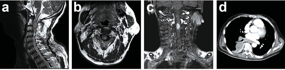
A 62-year-old male presented with a one-month history of progressive weakness of his right limbs, accompanied with pain of occipitocervical region. Physical examination revealed a severe right hemiplegic paralysis and mild urinary incontinence. MRI sagittal T2 images showed a slightly high signal of occupancy within the spinal cord at C2/C3 level, which was covered with an arc-shaped low signal at the superior and inferior margins (Figure 1a). Contrast-enhanced MRI images showed a markedly inhomogeneous enhancement at C2/C3 level (Figure 1b, 1c). Moreover, during the period of routine preoperative examination, thoracic CT unexpectedly discovered a huge mass in right lower lobe and perihilar without any cancer-related clinical symptoms were noted (Figure 1d). Then, the patient was examined by whole-body positron emission tomography (PET-CT) which showed an enormous tumor in lung and an isolated intramedullary spinal cord neoplasm, no additional systemic metastasis were found. A intramedullary tumor resection was performed.
NEUROPATHOLOGY
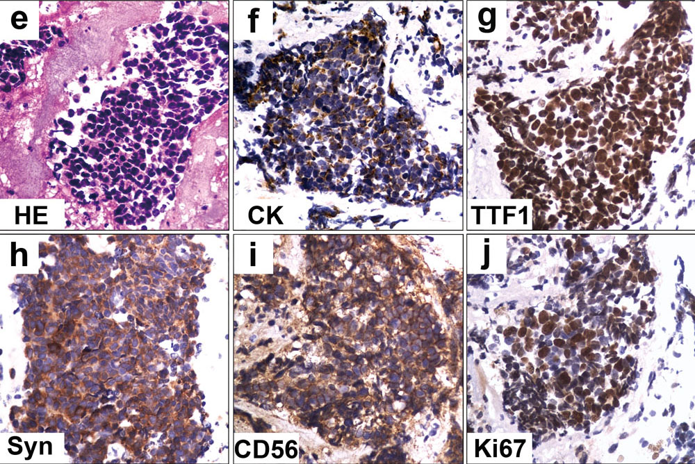
Histopathological examination revealed a infiltrated neoplasm in spinal cord, which consisted of sheets of densely packed small round or oval cells with hyperchromatic nuclei and little eosinophilic cytoplasm (HE, 400X, Figure 1e). Immunohistochemical examination showed positivity for CK (Pan-keratin, 400X, Figure 1f), TTF1 (400X, Figure 1g), Syn (400X, Figure 1h) and CD56 (400X, Figure 1i). The positive staining rate of Ki67 was 80% (400X, Figure 1j)
What is your diagnosis?