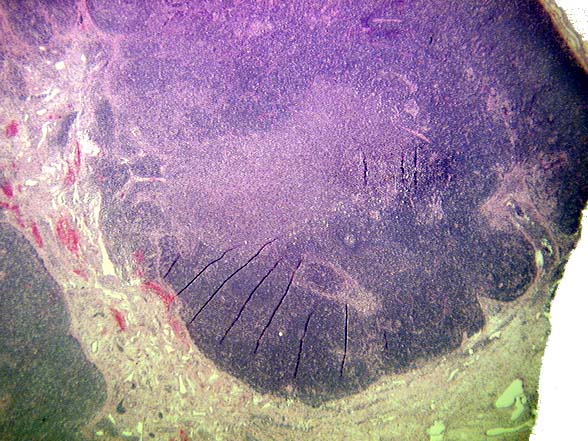
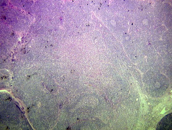
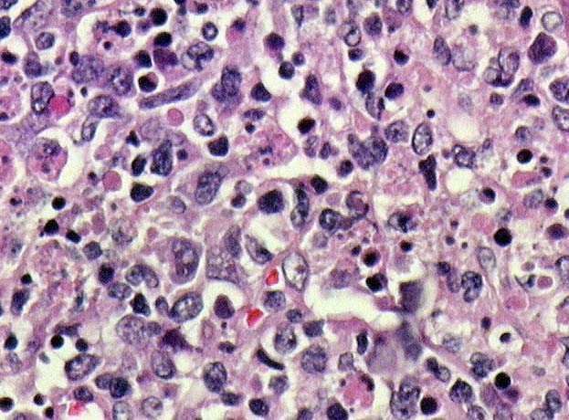
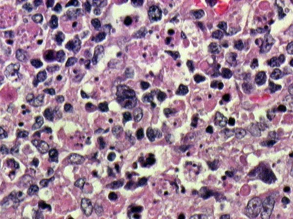
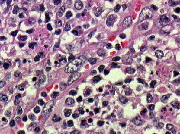
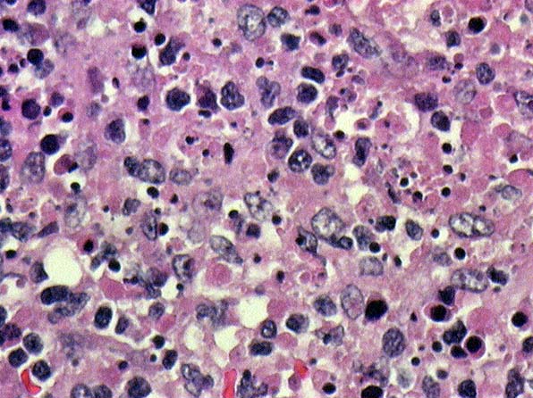
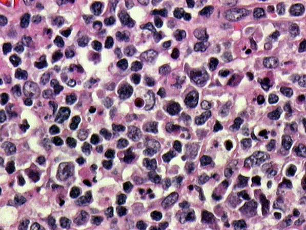
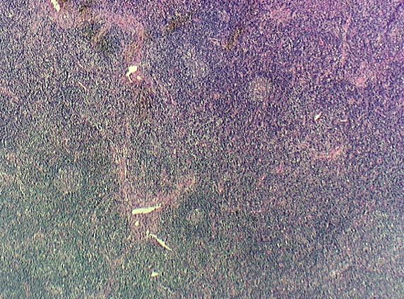
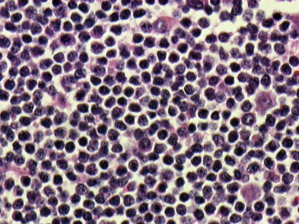

MICROSCOPIC DESCRIPTION:
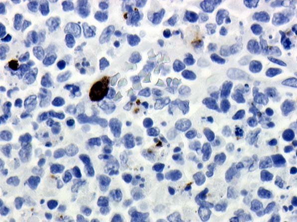
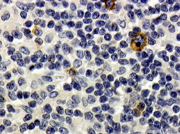
Histologic sections demonstrated multiple, discrete, varying sized, pale staining foci distributed throughout a lymph node. These foci consisted of histiocytes, small and some transformed lymphoid cells, and abundant karyorrhectic debris. No neutrophils were identified in these areas (Images 01, 02, 03, 04, 05, 06 and 07). Follicles, some with reactive changes, prominent T-zone nodules, pigment and an interfollicular expansion were also present (Images 08 and 09).
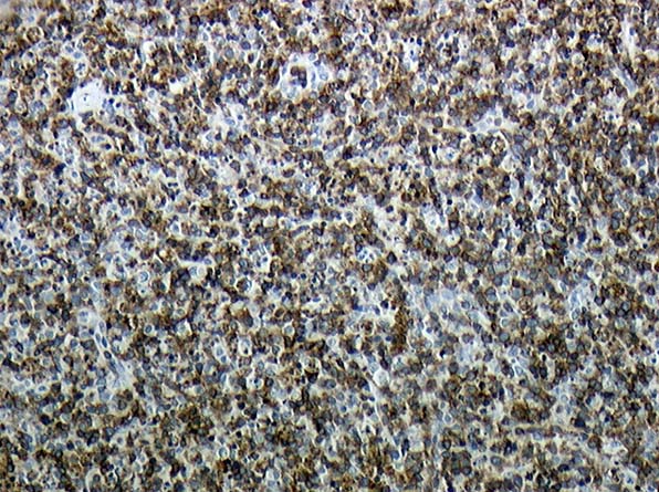
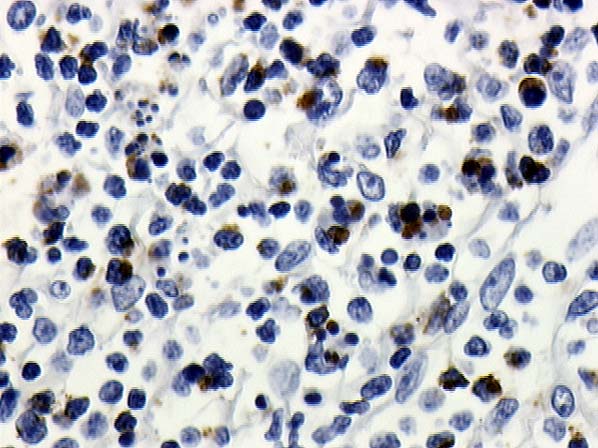
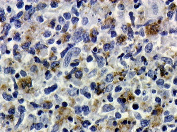
The following immunohistochemical stains were performed:
| Antigen/antibody | Usual reactivity | Result |
|---|---|---|
| LCA | Leukocytes | Numerous positive cells. |
| CD20/L26 (Image 10). | B cell | Highlights follicles and scattered positive cells of variable size including some very large forms, more numerous in some areas. |
| CD3 (Image 11) | T cells | Numerous positive cells |
| TIA 1 (Image 12) | Cytolytic T cells, NK cell subset | Moderate amount of positivity in areas with necrosis |
| CD68/PGM-1 (Image 13) | Monocyte, macrophage, some lymph | Moderate number of positive cells in areas of necrosis plus scattered positive cells throughout lymph node |
| CD15/Leu M1 (Image 14) | Reed-Sternberg, myeloid | Rare large cells positive, probably histiocytes |
| CD30/Ber H2 (Image 15) | Reed-Sternberg, activated lymph | Scattered positive large cells |