MICROSCOPIC DESCRIPTION:
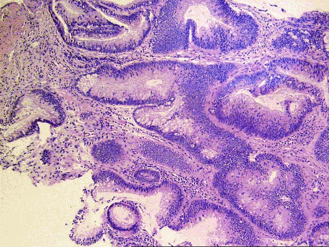
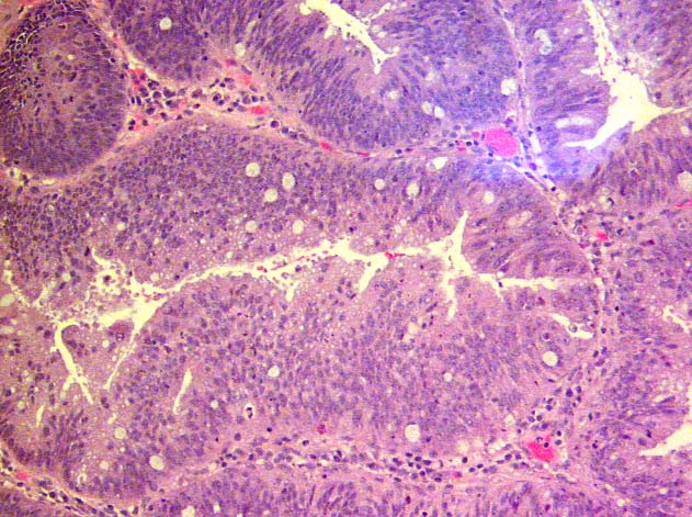
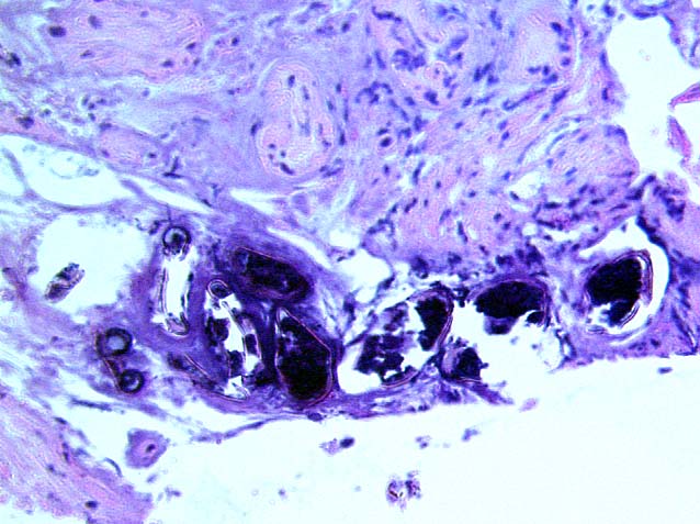
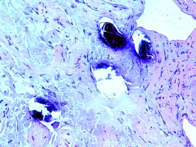
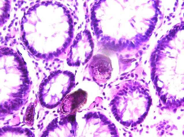
Histologic sections of rectal mucosa demonstrate a focus of adenomatous mucosa with focal moderate dysplasia. No invasive carcinoma is identified. However, widespread submucosal and occasional intramucosal calcified bodies are noted in many sections of this specimen. These bodies are often clustered, and elicit only a very focal mild inflammatory infiltrate, without granulomatous or giant cell reaction. The majority of involved areas demonstrate dense fibrosis without inflammation. Many of the bodies are in various states of breakdown and degradation, which may be due to chronic inflammatory reaction or may be an artefact of tissue processing.
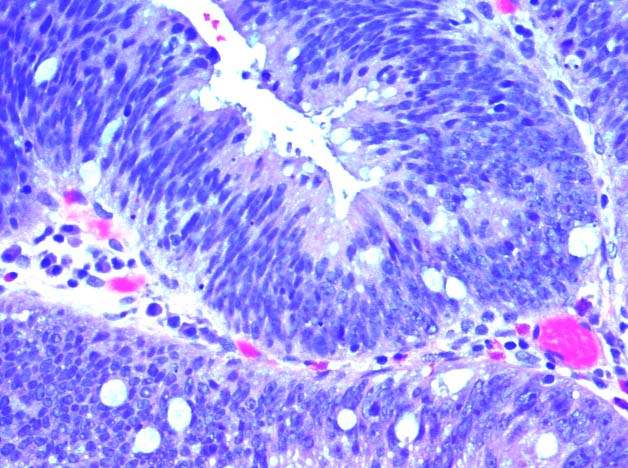
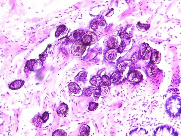
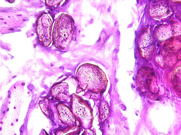
Careful examination of the better preserved calcified bodies on higher power demonstrate uniform oval shape and little variation in size. The bodies display a thick wall surrounding each structure, with no or little variation in the wall thickness. The bodies display a distinct internal structure with round to granular bodies of varying sizes and staining characteristics. Examination of the external surface of several of the bodies reveals occasional forms suggestive of spines or hooks which appear terminal or slightly lateral. No well-defined spines are discernible due to the advanced calcification of the bodies.
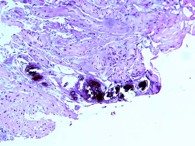
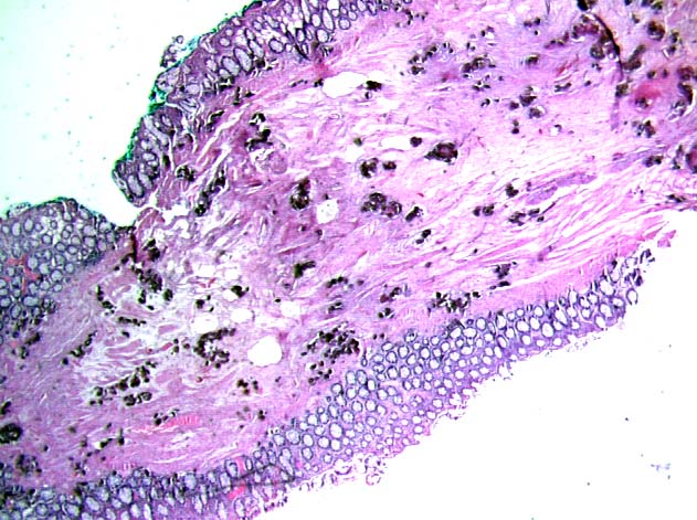
Identification of these bodies in the current tissue sample prompts review of the original carcinoma resection. This specimen displays a well- to moderately differentiated colonic adenocarcinoma, arising in a tubular adenoma with multifocal high grade dysplasia. Careful examination of the submucosa in several of the tumor fragments again reveals clustered refractile bodies with calcification, identical in appearance to those noted in the current tissue.
|
|
![]()
![]()
![]()
![]()