RADIOLOGY:
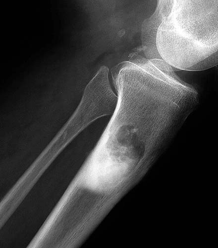
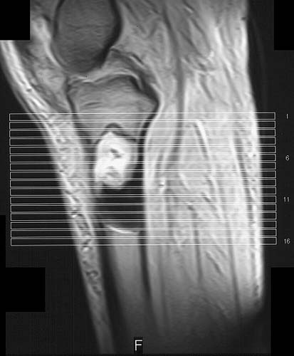
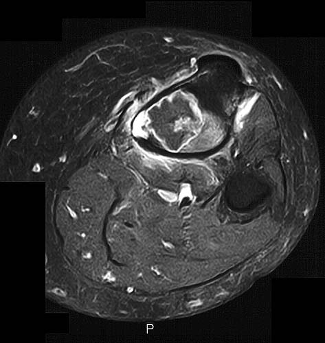
The plain films and CT scan (1 - 2) demonstrate a mixed lytic and sclerotic lesion of the proximal medial tibia with cortical destruction medially. There is a small soft tissue mass closely approximating this bone lesion. The lesion does not involve the adjacent fibula, patella or femur.