


MICROSCOPIC DESCRIPTION:
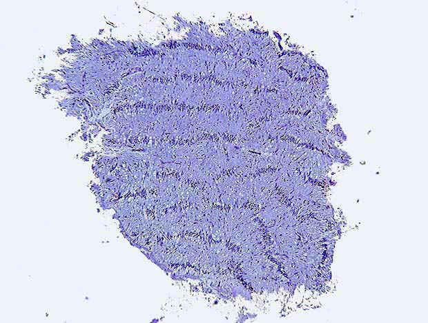
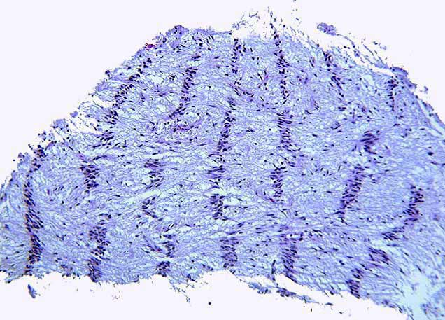
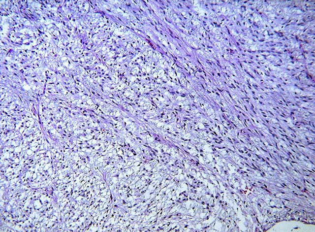
Sections of the first resection showed a fibrillary tumor with a distinctive palisading pattern in many of the fragments (Image 1,
Image 2,
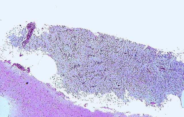
Image 3). In other areas this pattern was lost and the tumor had microcystic features with overall low cellularity
(Image 4). In some areas there was juxtaposition of the palisading pattern and a fibrillary pattern
(Image 5). There were no mitoses.
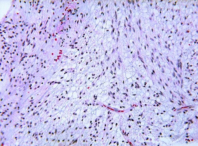
The second resection showed only rare areas of palisading cells (Image 6). Most of the tumor now showed a loosely cellular astrocytic neoplasm with prominent microcystic changes alternating with more fibrillar areas (Image 7 and Image 8). Again, there were no mitoses.