

MICROSCOPIC DESCRIPTION:
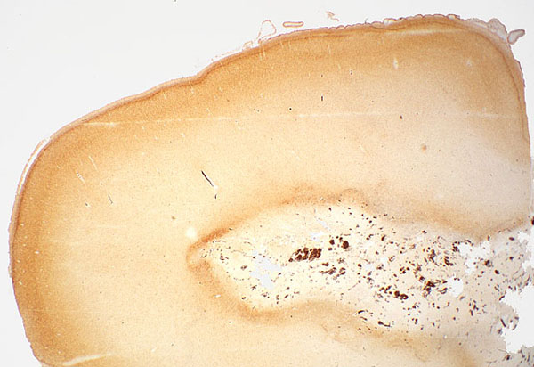
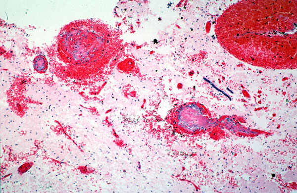
In a Bielschowsky preparation, the necrotic deep white matter was clearly demarcated by its pale appearance, and by a surrounding rim of brown/black argyrophilic debris. Veins in necrotic areas such as the thalamus were surrounded by petechial hemorrhages and contained organizing thrombi. Endothelialization of thrombi was clearly visible, e.g. in the germinal matrix. The choroid plexus was hemorrhagic, and the ventricles contained clotted blood. The vein of Galen and straight sinus were distended and engorged by large organizing thrombi, showing typical lines of Zahn.