

MICROSCOPIC DESCRIPTION:
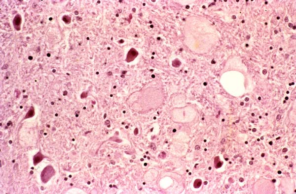
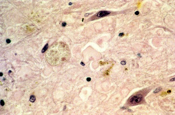
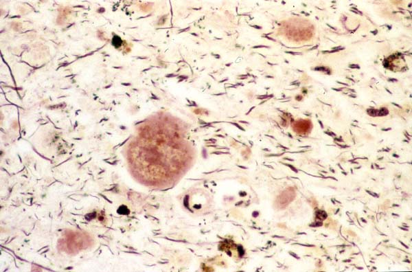
Fig. 3 shows numerous dystrophic axons in the globus pallidus with few residual neurons undergoing acute necrosis. Fig 4 is a high power view of the dystrophic axons, some of which contain hemosiderin pigment; few residual neurons are also seen. Prussian-blue stain (Fig. 5) highlights the abundant iron deposition in the substantia nigra which also displays dystrophic axons and near-total loss of neurons. Fig. 6 is a high-power view of Fig. 5. Bodian stain (Fig. 7) is negative in the large axonal spheroids.