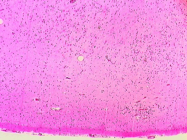
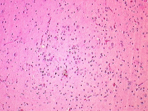

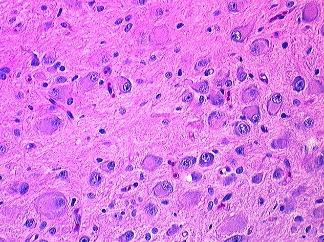
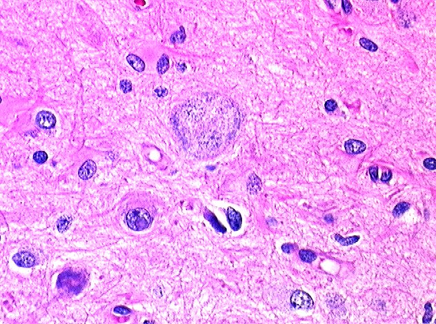
H&E stained sections reveal that at low power (40x) there is no obvious increase in cellularity and that the tissue resembles normal brain parenchyma. At slightly higher magnification, the numerous ganglionic neurons become apparent. In some areas, white matter tracts appear to be coursing through the hamartomatous nodule. The great variety in the size and shape of the neurons is better appreciated at higher magnification (200x). Binucleate ganglionic cells were extremely rare and in this micrograph, it is difficult to see both nuclei, although the Nissel substance is clearly visible as increased peripheral basophilia.