
MICROSCOPIC DESCRIPTION:
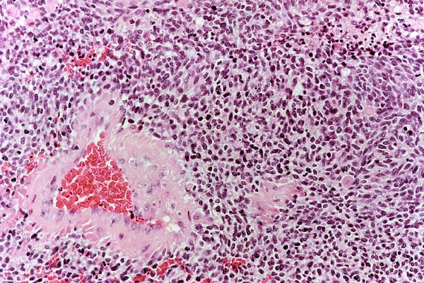
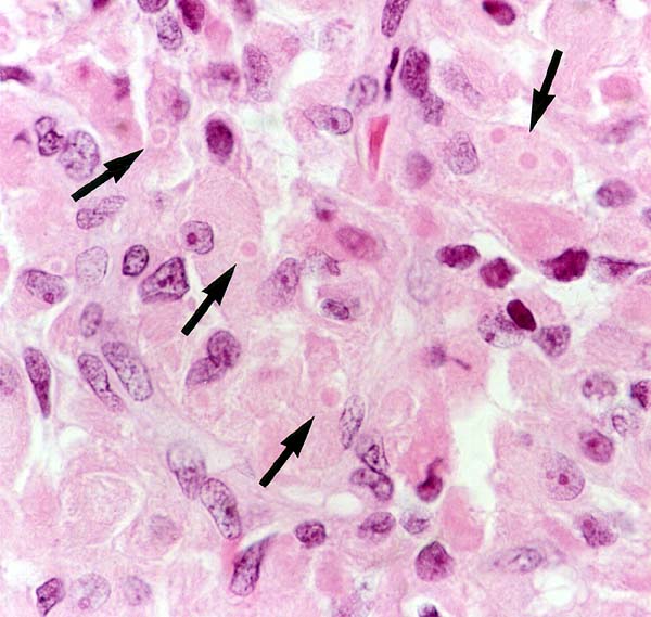
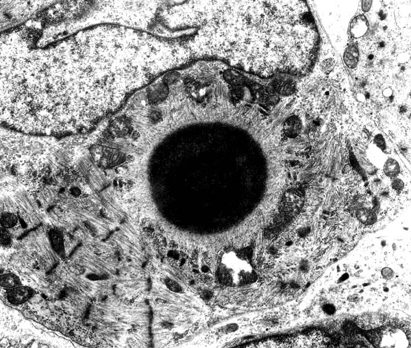
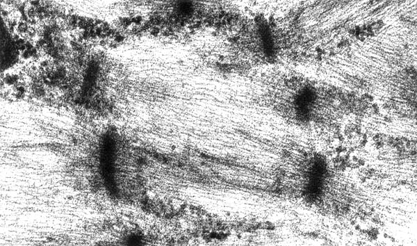
The histology revealed a markedly cellular tumour with a high mitotic rate, zonal necrosis, and hemorrhages (Figure 2). Scattered and in groups, cells with abundant bright eosinophilic cytoplasm stood out against a background of densely packed small oval or elongated cells with darkly staining hyperchromatic nuclei and indistinct cytoplasm. The identification in many of the large cells with cytoplasmic inclusions bodies consisting of a round eosinophilic core surrounded by a clear halo suggested a specific diagnosis (Figure 3, oil immersion). Immunostaining for synaptophysin and vimentin was negative. Focal immunopositivity for GFAP in the vicinity of necrosis and blood vessels was interpreted as native tissue frame. Large cells stained with antibodies to desmin and myoglobin (Figure 4). Ultrastructural studies are shown in Figure 5 (x7100) and Figure 6 (x25200). MYCN gene amplification was not demonstrable following FISH analysis and RNA PCR revealed no chromosomal translocation.