Brain Pathology Case of the Month - December 2017
FINAL DIAGNOSIS
Multiple intracranial tuberculomas.
DISCUSSION
Tuberculosis (TB) continues to be one of the important infectious disease worldwide, with more than 2 billion people affected (46, 102). Besides the pulmonary system, TB affects nearly every system in the body, including the lymph nodes, central nervous system (CNS) and others (133). Central nervous system tuberculosis remains one of the most severe manifestations of the disease(117). It can present in many forms, one of which is intracranial tuberculomas (ICT) which develops in 16-40% of affected patients (140, 150, 196).
We searched the PubMed for cases of multiple ICT, using various combinations of keywords including multiple intracranial tuberculomas, disseminated intracranial tuberculomas, multiple intracerebral tuberculomas, disseminated intracerebral tuberculomas, multiple brain tuberculomas, disseminated brain tuberculomas, brain tuberculomas, and trabecular brain abscess. A total of 387 abstracts were collected. Only articles with descriptive cases were filtered and all review articles were excluded. An attempt to retrieve the full-text articles through Google Scholar, King Hussein Cancer Center library, the library at the American University of Beirut and other sources was undertaken. Eventually a total of 162full text articles were collected. Additionally some clinical data could be retrieved from 3abstracts(2-9, 11-15, 17-19, 21-23, 25-27, 29-32, 35-41, 43, 44, 48-53, 56-65, 67-75, 77-99, 101, 103-105, 107-115, 118-127, 129, 135-137, 141, 142, 144, 145, 147-149, 151, 153, 155-157, 159-163, 167, 168, 170-176, 179-195, 198-209). The data then was summarized on an excel spread sheet and collected items included the author(s), publishing journal, the geographic location(based on the address of the corresponding author), age, gender, presenting symptoms and signs, radiological features, involvement of spine, the presence of other potential predisposing medical conditions, the presence of other sites affected by TB including lung, any previous or concurrent TB medications, and the outcome. Additionally the method of confirming the diagnosis such as biopsy, the use of special stains like ZN special stain and/ or polymerase chain reaction (PCR) were all collected.

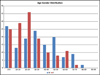
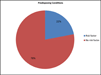
A total of 291 cases were retrieved including our own case. More than half of the reported cases came from Asia (n=155, 53.2%) (Figure- 2A). The male (n=136) and female (n=140) were affected almost equally with a male to female ratio of 1:1.03. The average age at diagnosis was 30.25 years, and around two thirds of the cases (n=192, 65%) were reported in the first 4 decades. Twenty percent of cases occurred in the 20-29 age group, which was the most common age group affected. Pediatric patients (0-19 years of age) constituted one third of the affected individuals (n=95, 33.8%). Females were more likely to be affected in 2ndand3rddecades, while males were more likely to be affected in later decades (Figure-2B).
Symptoms of increased intracranial pressure were the most common presenting manifestations. Headache was the most common presenting symptom (n=108, 37.1%), followed by fever (n=83, 28.5%). Paresis (n=84, 28.9%) was the most common sign, followed by altered mental status in 64 (21.9%) patients. Many patients however; presented with more than one complaint and the combination of headache, fever, and weakness was the most common.
The lesions were mostly seen in the cerebrum involving the frontal lobe in 50(17.2%) cases, the parietal lobe in 41 (14%), the temporal lobe in 34 (11.7%), and the occipital lobe in 22 (7.6%). This was followed by the cerebellum in 97 (33.3%) cases, brain stem in 22 (7%), including the pons in 16 (5%), thalamus in 22 (7%), and the basal ganglia in 12 (4%). Radiologically; enhancing multiple lesions was the most common pattern (n=288, 99%). The enhancement was heterogonous in 2 (0.7%) cases and in one case (0.3%) the lesions were non-enhancing. Edema (n=65, 22%) and hydrocephalus (n=30, 10%) were reported in few cases as well as infarction (n=10, 3%). Leptomeningeal enhancement was seen in 14(5%) cases only. Spinal involvement was reported in only a minority of cases (n=23, 8%).
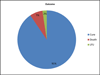
Concurrent or previous pulmonary involvement was reported in around a third of cases (n=106, 36%). Predisposing conditions were reported in another quarter of cases (n=64, 22.6%) These conditions included HIV (n =10),use of immunosuppressant agents (n=10), malignancies (n=8), diabetes mellitus (n=4), alcoholism (n=4), systemic lupus erythematosus (SLE) (n=3), liver cirrhosis (n=3), and malnutrition (n=2). Interestingly pregnancy was reported in 6 cases. History of contact with tuberculosis patients (n=21) was also a potential important predisposing condition (Figure 2C).
Diagnosis was confirmed by a tissue biopsy in 41% (n=118) of cases, including postmortem tissue diagnosis (n=3) as well as abscess aspiration(n=12). Granulomatous inflammation either caseating or non-caseating was seen. The ZN special stain was performed in 131 (45%) cases and was positivein70(53%) cases, while it was negative in 61 (47%). The PCR was performed in 57(19%) cases, and was positive in 30 (52.6%)cases, while it was negative in 27 (47.4%).
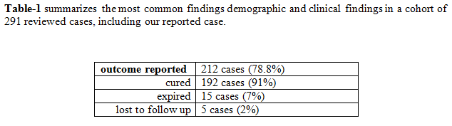
Treatment was documented in only 220 (75.0%). Of these anti-tuberculous medication was used alone in 79% (n=174), medication and surgery in 18% (n=40), and surgical alone in 3% (n=6). The outcome was reported in 212 (72.8%) cases. Most cases (n=192, 91%) were cured either completely or partially. Death was reported in only 7% (n=15) of cases, the rest (n=5, 2%) were lost to follow up (Figure 2D).
We are presenting a case of multiple ICT along with an extensive literature review on the topic. CNS tuberculosis accounts for approximately 1% of all cases of tuberculosis(146), and about 10% of patients with tuberculosis develop CNS disease (47). The presence of an active pulmonary tuberculosis on chest X-ray ranges from 30 to 50% in a recent series (166, 178). CNS involvement is noted in 5 to 10% of extra-pulmonary tuberculosis(143), however, most multiple ICT have extra-CNS tuberculosis(1). CNS involvement by tuberculosis has 3 different forms including meningitis, tuberculomas and spinal arachnoiditis. Intracranial tuberculomas are the least common presentation, reported in 1% of these patients (128), and they are multiple in only 15%-33% of the cases (66). While tuberculous meningitis affects mainly children, tuberculoma and spinal arachnoiditis are seen mainly in adults (10).Intracranial tuberculoma has a low incidence in developed countries (0.15 to 0.18%), but remains a challenging condition in developing countries, as it can reach up to 15 - 30% of brain tumorous lesions (28). Interestingly; around 2/3 (66.0%) of the summarized cases in this cohort presumably originated from Asia and Africa. Multiple ICT affected younger age groups and one third of cases affected the pediatric age groups (0-19 years).
The two main routes of infection in CNS tuberculosis are the hematogenous, which is the most common, and the direct spread. The hematogenous pathway is proposed to be a two-stage process. First tuberculous lesions (Rich's focus) develop in the brain during the stage of bacteremia or shortly afterward. Later on rupture or growth of one or more of these lesions leads to the development of CNS tuberculosis. Eventually, the location of TB lesions is directly related to the pattern of blood flow. It usually spreads to the cerebral hemispheres and basal ganglia in adults and to the cerebellum in children (42).These initial tuberculous lesions may be located in the meninges, the subpial or subependymal surfaces of the brain or in the spinal cord, and may remain dormant for years after initial infection. The causes and the mechanisms that control this distribution are still unknown. The direct spread, on the other hand, usually occurs into the meninges from the middle ear or the calvarial bone(33). It is presumed that multiple IC tuberculomas are mostly secondary to hematogenous spread, while the solitary IC ones evolve from CSF infection into the adjacent parenchyma (1).
When comparing solitary versus multiple ICT several differences become apparent. There is a cleat predominance of solitary ICT with a M:F ratio of 1:3.5(1), in contrast to the almost equal distribution in males and females in multiple ICT. However; other reports show an almost equal distribution between both genders (134). Furthermore; multiple ICT appear to affect younger patients. The median age for solitary IC ranges from 38-42 years (134), while the median age in our review is of 28 years. This has practical implications. The presence of multiple IC lesions in a young patient should warrant consideration of infections over metastatic carcinoma, however, in older age groups the reverse should be considered. Interestingly, most cases with multiple IC tuberculomas lacked a clear predisposing condition that might affect the immunity, as less than a quarter of cases (22.6%)had predisposing conditions. These included HIV infection, malnutrition, alcoholism, other malignancies, the use of immunosuppressant agents(146),systemic lupus erythematosus (SLE)(132), diabetes mellitus (DM)(20), liver cirrhosis (100, 177), and pregnancy (16). History of contact with other tuberculous patients was reported in some patients (24). Once the diagnosis is established the outcome is favorable in both conditions. 75.8% of all cases in the single IC recovered completely, while death was reported in 4% (134). In another study, the cure rate was 40%, partial recovery was 40%, and death was 6% (1) . In our literature review 64% of multiple IC tuberculomas showed complete recovery, 32% showed partial recovery and the reported death rate was 4%.
Metastatic malignancy remains the most common cause for multiple IC ring-enhancing lesions (154). However, in developing countries infectious diseases appear to be encountered more frequently(55), and remains an important consideration, especially in young individuals. While tuberculosis remains the most common infectious pathology, neurocysticercosis and toxoplasmosis should be ruled out (55) . Neurocysticercosis (NCC) is caused by the larval stage of Taenia solium, is the most common and serious parasitic disease of the CNS in many developing countries (76).The usual mode of presentation is focal seizures with or without secondary generalization, in a previously healthy individual (164), reported in up to 79% of neurocysticercosis cases (197). Other complaints are related to space occupying lesions including headache, vomiting (169) and features of raised intracranial pressure(165). This contrasts with the mode of presentation of IC tuberculomas in which complaints related to increased IC pressure predominate. Seizure was reported in only 19.8% of our cases. The presence of subcutaneous nodules in neurocysticercosis usually confirms the diagnosis (54) . The characteristic appearance of neurocysticercosis in MRI include single, which is more common, or multiple enhancing lesions with diameter range between 5-20mm in any part of cerebral hemispheres. 44% of patients have punctuated eccentric high density structure suggestive of scolex. Another minor characteristic finding of neurocysticercosis is rounded hypotense non-enhancing lesions (123) . The radiological appearance of IC tuberculoma in MRI is hypointensity with ring-shaped contrast enhancement on T1-weighted images, and hyperintensity with a central hypointensity on T2-weighted images(196) .
Toxoplasmosis, on the other hand, is a protozoan infection and is one of the most common causes of focal brain lesions in patients with Human Immunodeficiency virus/acquired immune deficiency syndrome (HIV/AIDS) particularly in developing countries (152, 158) . CT and MR images usually show multiple ring-enhancing lesions (139) which is similar to multiple intracranial tuberculosis. The characteristic imaging pattern in toxoplasmosis lesions however, is a ring-enhancing lesion made up of an eccentric nodule. In around 30% of the lesions, this enhancing nodule is found within and adjacent to the enhancing rim (138) Most lesions in toxoplasmosis occur in basal ganglia (130, 131) and in frontal and parietal lobes(116, 130), which is different from the distribution of the cases in multiple IC tuberculomas.
Another important consideration in multiple IC tuberculomasis differentiating it from metastatic malignancy. Besides clinical history, radiology remains an important techniques that can be used nowadays, through measuring the regional cerebral blood flow (rCBV) which is typically elevated in malignancies but not in tuberculomas(34) . The definitive diagnostic modality however remains to obtain a tissue diagnosis. This is a risky procedure that might be associated with complications, especially brain hemorrhage, which occur in 1.3% to 5% of cases (106). In the absence of a history of malignant tumor, a presumptive anti-tuberculous therapy may be proposed as a diagnostic and therapeutic test (45).
In conclusion, we present a case of multiple ICT in a young female patient, with a previous history of pulmonary TB. Multiple ICT remain diagnostically challenging and sorting out the differential diagnosis with other conditions including other infections and metastatic malignancy is crucial. Multiple ICT predominates in less developed countries, and although males and females are affected almost equally, females predominate in younger age groups. Around one third of all reported cases affected children. Spinal involvement is rare and the presence of predisposing conditions can be seen in around a quarter of cases. Establishing the correct diagnosis is of utmost importance since the outcome is favorable with the appropriate management.
Acknowledgments
The authors would like to thank Dr. Nader Hermas for his help in getting the articles from the medical library of the American University of Beirut.
REFERENCES
- Abuhamed MM, Bo X, Xiaoqin L, Fufeng Z, Long L, Fangfang B, Jing L (2009) Comparison between solitary and multiple intracranial tuberculoma. Neurosciences (Riyadh).14(3):254-9.
- Adiego MI, Millan J, Royo J, Dominguez L, Castellote MA, Alfonso JI, Valles H (1994) Unusual association of secondary tonsillar and cerebral tuberculosis. J Laryngol Otol.108(4):348-9.
- Afghani B, Lieberman JM (1994) Paradoxical enlargement or development of intracranial tuberculomas during therapy: case report and review. Clin Infect Dis.19(6):1092-9.
- Ahmadi F, Nashibi R, Naghieh M, Feizi J, Shirmardi M (2014) Multiple brain tuberculomas in a 32-year-old woman with chronic headache. Arch Iran Med.17(10):724-5.
- Ahmetgjekaj I, Kabashi-Mucaj S, Lascu LC, Bondari S, Bondari A (2014) Paradoxical Growth of Optochiasmatic Tuberculoma during the Treatment of Tuberculous Meningitis. Curr Health Sci J.40(3):225-7.
- Ahn JS, Yang DH, Kim YK, Cho SH, Kim IY, Lee JJ, Chung IJ, Kim HJ (2007) Multiple intracranial tuberculomas mimicking granulocytic sarcomas in acute myeloid leukemia. J Korean Med Sci.22 Suppl:S171-3.
- Ahn JY, Chang JH, Kim KS, Kim WJ (2007) Disseminated tuberculosis with multiple intracerebral tuberculomas in a patient with anorexia nervosa. Int J Eat Disord.40(3):288-91.
- Akhaddar A, Boucetta M (2011) Images in clinical medicine. Multiple intracranial tuberculomas. N Engl J Med.365(16):1527.
- Akritidis N, Galiatsou E, Kakadellis J, Dimas K, Paparounas K (2005) Brain tuberculomas due to miliary tuberculosis. South Med J.98(1):111-3.
- al-Deeb SM, Yaqub BA, Sharif HS, Motaery KR (1992) Neurotuberculosis: a review. Clin Neurol Neurosurg.94 Suppl:S30-3.
- Al-Nozha M, Naim Ur R, Akhtar J (1986) Intracranial tuberculoma: case report and review of the literature. Trop Geogr Med.38(4):425-8.
- Alame T, Keller K, Michel O, Sergysels R (1996) Hyperthermia occurring with paradoxical development of cerebral tuberculomas. Respiration.63(6):381-3.
- Alarcon F, Maldonado JC, Rivera JW (2011) Movement disorders identified in patients with intracranial tuberculomas. Neurologia.26(6):343-50.
- Alkan A, Parlak M, Baysal T, Sigirci A, Kutlu R, Altinok T (2003) En-plaque tuberculomas of tentorium in a pregnant woman: follow-up with MRI(2003:2b). Eur Radiol.13(5):1190-3.
- Alkhani A, Al-Otaibi F, Cupler EJ, Lach B (2006) Miliary tuberculomas of the brain: case report. Clin Neurol Neurosurg.108(4):411-4.
- Arseni C (1958) Two hundred and one cases of intracranial tuberculoma treated surgically. J Neurol Neurosurg Psychiatry.21(4):308-11.
- Artopoulos J, Chalemis Z, Christopoulos S, Manios S, Kelekis L (1984) Sequential computed tomography in tuberculous meningitis in infants and children. Comput Radiol.8(5):271-7.
- Awada A, Daif AK, Pirani M, Khan MY, Memish Z, Al Rajeh S (1998) Evolution of brain tuberculomas under standard antituberculous treatment. J Neurol Sci.156(1):47-52.
- Bahemuka M, Murungi JH (1989) Tuberculosis of the nervous system. A clinical, radiological and pathological study of 39 consecutive cases in Riyadh, Saudi Arabia. J Neurol Sci.90(1):67-76.
- Baker MA, Lin HH, Chang HY, Murray MB (2012) The risk of tuberculosis disease among persons with diabetes mellitus: a prospective cohort study. Clin Infect Dis.54(6):818-25.
- Bas NS, Guzey FK, Emel E, Alatas I, Sel B (2005) Paradoxical intracranial tuberculoma requiring surgical treatment. Pediatr Neurosurg.41(4):201-5.
- Basoglu OK, Savas R, Kitis O (2002) Conventional and diffusion-weighted MR imaging of intracranial tuberculomas. A case report. Acta Radiol.43(6):560-2.
- Basta M, Lydakis C, Daskalogiannaki M, Schiza S, Siafakas NM (2001) Multi-focal tuberculosis with multiple intracranial tuberculomas in a non-immunocompromised patient. Respir Med.95(10):841-3.
- Becerra MC, Appleton SC, Franke MF, Chalco K, Arteaga F, Bayona J, Murray M, Atwood SS, Mitnick CD (2011) Tuberculosis burden in households of patients with multidrug-resistant and extensively drug-resistant tuberculosis: a retrospective cohort study. Lancet.377(9760):147-52.
- Berger P, Larson J, Guss D (1998) Central nervous system tuberculoma: a case report. J Emerg Med.16(5):719-22.
- Bhargava S, Tandon PN (1980) Intracranial tuberculomas: a CT study. Br J Radiol.53(634):935-45.
- Bishburg E, Sunderam G, Reichman LB, Kapila R (1986) Central nervous system tuberculosis with the acquired immunodeficiency syndrome and its related complex. Ann Intern Med.105(2):210-3.
- B�l�k A, T�rk �, Ar?ba? E, K�krek Z (1998) Intracranial tuberculoma: Clinical and MRI findings. Turgut ozal Tip merkezi dergisi.5(2):3.
- Brid NS, Kulkarni AG, Kale MM, Shah PJ, Yadav SR (1997) Ring-enhancing lesions on computed tomography. Postgrad Med J.73(860):355-6.
- Capon A, Noterman J, Hubert JP, Klastersky J, Flament-Durand J (1975) Multiple tuberculomas of the brain. Report of a case. Acta Neurochir (Wien).32(3-4):303-12.
- Ceylan E, Gencer M (2005) Miliary tuberculosis associated with multiple intracranial tuberculomas. Tohoku J Exp Med.205(4):367-70.
- Chang T, Rodrigo C, Ranawaka N, Atukorala I (2012) Multiple ring-enhancing cerebral lesions in systemic lupus erythematosis: a case report. J Med Case Rep.6:172.
- Chatterjee S (2011) Brain tuberculomas, tubercular meningitis, and post-tubercular hydrocephalus in children. J Pediatr Neurosci.6(Suppl1):S96-S100.
- Chatterjee S, Saini J, Kesavadas C, Arvinda HR, Jolappara M, Gupta AK (2010) Differentiation of tubercular infection and metastasis presenting as ring enhancing lesion by diffusion and perfusion magnetic resonance imaging. Journal of Neuroradiology.37(3):167-71.
- Chatterjee S, Saini J, Kesavadas C, Arvinda HR, Jolappara M, Gupta AK (2010) Differentiation of tubercular infection and metastasis presenting as ring enhancing lesion by diffusion and perfusion magnetic resonance imaging. J Neuroradiol.37(3):167-71.
- Chaudhry LA, Ebtesam B-E, Al-Solaiman S (2012) Milliary tuberculosis with unusual paradoxical response at 3 weeks of antituberculous treatment. J Coll Physicians Surg Pak.22(1):43-5.
- Cho YH, Ho TS, Wang SM, Shen CF, Chuang PK, Liu CC (2014) Childhood tuberculosis in southern Taiwan, with emphasis on central nervous system complications. J Microbiol Immunol Infect.47(6):503-11.
- Chou PS, Liu CK, Lin RT, Lai CL, Chao AC (2012) Central nervous system tuberculosis: a forgotten diagnosis. Neurologist.18(4):219-22.
- Coulter JB, Baretto RL, Mallucci CL, Romano MI, Abernethy LJ, Isherwood DM, Kumararatne DS, Lammas DA (2007) Tuberculous meningitis: protracted course and clinical response to interferon-gamma. Lancet Infect Dis.7(3):225-32.
- Danziger A, Morehouse HT, Horoupian DS (1981) CT findings in multiple tuberculomata. Comput Tomogr.5(1):69-71.
- Darwish B, Blackmore T, Hunn M (2001) Intracranial and spinal tuberculosis requiring neurosurgical intervention. The Wellington Hospital experience 1998-2001. N Z Med J.114(1141):445-7.
- Dastur DK, Lalitha VS, Prabhakar V (1369) Pathological analysis of intracranial space-occupying lesions in 1000 cases including children. 1. Age, sex and pattern; and the tuberculomas. J Neurol Sci.6(3):575-92.
- Dastur HM, Desai AD (1965) A comparative study of brain tuberculomas and gliomas based upon 107 case records of each. Brain.88(2):375-96.
- de la Riva P, Urtasun M, Castillo-Trivino T, Camino X, Arruti M, Mondragon E, Lopez de Munain A (2013) Clinical response to thalidomide in the treatment of intracranial tuberculomas: case report. Clin Neuropharmacol.36(2):70-2.
- Del Brutto OH, Mosquera A (1999) Brainstem tuberculoma mimicking glioma: The role of antituberculous drugs as a diagnostic tool. Neurology.52(1):210.
- Dheda K, Barry CE, 3rd, Maartens G (2015) Tuberculosis. Lancet.
- Dye C, Scheele S, Dolin P, Pathania V, Raviglione MC (1999) Consensus statement. Global burden of tuberculosis: estimated incidence, prevalence, and mortality by country. WHO Global Surveillance and Monitoring Project. JAMA.282(7):677-86.
- el-Sonbaty MR, Abdul-Ghaffar NU, Marafy AA (1995) Multiple intracranial tuberculomas mimicking brain metastases. Tuber Lung Dis.76(3):271-2.
- Ersahin M, Hakan T, Ayan E, Berkman Z, Ekinci O, Ceran N, Aker FV (2010) Diagnostic and therapeutic role of CT-guided stereotactic surgery in the management of intracranial tuberculomas. Turk Neurosurg.20(3):295-302.
- Etienne M, Noble JM (2007) Multiple parenchymal tuberculomas without tuberculous meningitis. Arch Neurol.64(7):1045-7.
- Fan ZM, Zeng QY, Huo JW, Bai L, Liu ZS, Luo LF, Yang JC, Zhou XH (1998) Macronodular multi-organs tuberculoma: CT and MR appearances. J Gastroenterol.33(2):285-8.
- Gamage R, Seneviratne U, Constantine GR (2000) Paradoxical progression of intracranial tuberculous lesions during treatment. Ceylon Med J.45(1):31.
- Garcia-Vidal C, Cabellos C (2008) Paradoxical reaction of multiple cerebral tuberculomas. Mayo Clin Proc.83(3):264.
- Garg R (2001) Medical management of neurocysticercosis. Neurol India.49(4):329-37.
- Garg RK, Desai P, Kar M, Kar AM (2008) Multiple ring enhancing brain lesions on computed tomography: An Indian perspective. J Neurol Sci.266(1-2):92-6.
- Garg RK, Kar AM, Kumar T (2000) Neurocysticercosis like presentation in a case of CNS tuberculosis. Neurol India.48(3):260-2.
- Garner J, Howard R (2013) Ring-enhancing lesions in the brain and spinal cord. BMJ Case Rep.2013.
- Gasparetto EL, Tazoniero P, de Carvalho Neto A (2003) Disseminated tuberculosis in a pregnant woman presenting with numerous brain tuberculomas: case report. Arq Neuropsiquiatr.61(3b):855-8.
- Golub V, Malhotra P, Patel S (2011) Mycobacterial brain tuberculomas due to Bacille Calmette-Guerin intravesical chemotherapy for bladder cancer: A case report and literature review. Can J Infect Dis Med Microbiol.22(3):104-6.
- Gondal M, Mushtaq S, Hussain T (2013) Brain tuberculomas mimicking intracranial metastasis in a patient presenting with fits. J Coll Physicians Surg Pak.23(10):748-9.
- Gropper MR, Schulder M, Duran HL, Wolansky L (1994) Cerebral tuberculosis with expansion into brainstem tuberculoma. Report of two cases. J Neurosurg.81(6):927-31.
- Guest SS, Sivit CJ, Meisler WJ, Stevens AC, Simon GL (1987) Intracranial tuberculosis due to Mycobacterium bovis. Comput Radiol.11(3):151-4.
- Guzel A, Tatli M, Aluclu U, Yalcin K (2005) Intracranial multiple tuberculomas: 2 unusual cases. Surg Neurol.64 Suppl 2:S109-12.
- Hasan Kocaeli OT, Elif Basaran, Ahmet Karaoglu,, Bekar aA (2011) Multiple Dural Tuberculomas Presenting as Leptomeningeal Carcinomatosis. Case Reports in Neurological Medicine.2011:4.
- Heath PD, Grant JW (1984) Intracranial infection due to mycobacterium bovis in Hodgkin's disease. Br Med J (Clin Res Ed).288(6415):465-6.
- Hejazi N, Hassler W (1997) Multiple intracranial tuberculomas with atypical response to tuberculostatic chemotherapy: literature review and a case report. Acta Neurochir (Wien).139(3):194-202.
- Hejazi N, Hassler W (1997) Multiple intracranial tuberculomas with atypical response to tuberculostatic chemotherapy: literature review and a case report. Infection.25(4):233-9.
- Helmy A, Antoun N, Hutchinson P (2011) Cerebral tuberculoma and magnetic resonance imaging. J R Soc Med.104(7):299-301.
- Henderson C, Meyers B, Humayun Gultekin S, Liu B, Zhang DY (2003) Intracranial tuberculoma in a liver transplant patient: first reported case and review of the literature. Am J Transplant.3(1):88-93.
- Hildebrandt G, Agnoli AL (1982) Differential diagnosis and therapy of intracerebral tuberculomas. J Neurol.228(3):201-8.
- Ho BL, Hsu CY (2008) Miliary intracranial tuberculomas presenting as rapidly reversible encephalopathy. Acta Neurol Taiwan.17(2):149-50.
- Huang CR, Lui CC, Chang WN, Wu HS, Chen HJ (1999) Neuroimages of disseminated neurotuberculosis: report of one case. Clin Imaging.23(4):218-22.
- Idris MN, Sokrab TE, Arbab MA, Ahmed AE, El Rasoul H, Ali S, Elzubair MA, Mirgani SM (2007) Tuberculoma of the brain: a series of 16 cases treated with anti-tuberculosis drugs. Int J Tuberc Lung Dis.11(1):91-5.
- Iqbal N, Natarajan N, Periyasamy S, George S, Basheer A, Mookkappan S (2014) Miliary tuberculosis with left brachial monoplegia: A case report. Australas Med J.7(10):400-4.
- Ishiwada N, Tokunaga O, Nagasawa K, Ichimoto K, Kinoshita K, Hishiki H, Kohno Y (2013) Isoniazid- and streptomycin-resistant miliary tuberculosis complicated by intracranial tuberculoma in a Japanese infant. Tohoku J Exp Med.229(3):221-5.
- Ito A, Nakao M, Wandra T (2003) Human Taeniasis and cysticercosis in Asia. Lancet.362(9399):1918-20.
- Jain RS, Kumar S, Tejwani S (2015) A rare association of tuberculous longitudinally extensive transverse myelitis (LETM) with brain tuberculoma. Springerplus.4:476.
- ain V, Singhi P (2002) A "bunch of grapes" intracranial tuberculoma. Neurology.59(7):1111.
- Jain VK, Chandramukhi A, Venkataramana NK, Das BS, Gokul BN, Rao TV (1989) The far cry of a TB brain. Report of a case of tuberculous meningitis, multiple tuberculomas and tuberculous abscess. Clin Neurol Neurosurg.91(2):171-6.
- Janse van Rensburg P, Andronikou S, van Toorn R, Pienaar M (2008) Magnetic resonance imaging of miliary tuberculosis of the central nervous system in children with tuberculous meningitis. Pediatr Radiol.38(12):1306-13.
- Jorge JH, Graciela C, Pablo AP, Luis SH (2012) A life-threatening central nervous system-tuberculosis inflammatory reaction nonresponsive to corticosteroids and successfully controlled by infliximab in a young patient with a variant of juvenile idiopathic arthritis. J Clin Rheumatol.18(4):189-91.
- Kalita J, Misra UK (1998) EEG changes in tuberculous meningitis: a clinicoradiological correlation. Electroencephalogr Clin Neurophysiol.107(1):39-43.
- Kamble RB, N JP, Shivashankar R (2015) CT Perfusion Dynamics of Intracranial Tuberculomas. J Clin Diagn Res.9(5):Tc01-5.
- Kaminogo M, Ishimaru H, Morikawa M, Suzuki Y, Shibata S (2002) Proton MR spectroscopy and diffusion-weighted MR imaging for the diagnosis of intracranial tuberculomas. Report of two cases. Neurol Res.24(6):537-43.
- Kelly JD, Teeter LD, Graviss EA, Tweardy DJ (2011) Intracranial tuberculomas in adults: a report of twelve consecutive patients in Houston, Texas. Scand J Infect Dis.43(10):785-91.
- Kim DG, Lee JI, Lee DS, Lee MC, Choi KS, Han DH (1995) 99mTc-HMPAO labeled leukocyte SPECT in intracranial lesions. Surg Neurol.44(4):338-45.
- Kim KS, Weinberg PE (1982) Cerebral tuberculomas. Surg Neurol.18(3):203-5.
- Kim MS, Kim KJ, Chung CK, Kim HJ (2000) Intradural extramedullary tuberculoma of the spinal cord: a case report. J Korean Med Sci.15(3):368-70.
- Krygowski JD, Brennen DF, Counselman FL (2010) Intracranial tuberculomas: an unusual cause of altered mental status in a pediatric patient. J Emerg Med.38(3):323-7.
- Kumar R, Jain R, Kaur A, Chhabra DK (2000) Brain stem tuberculosis in children. Br J Neurosurg.14(4):356-61.
- Kumar S, Puri V, Mehndiratta MM, Gupta S, Bhutani A, Sharma C (1995) Paradoxical response to antitubercular drugs. Indian J Pediatr.62(6):695-701.
- Lebas J, Malkin JE, Coquin Y, Modai J (1980) Cerebral tuberculomas developing during treatment of tuberculous meningitis. Lancet.2(8185):84.
- Lee CH, Lui CC, Liu JW (2007) Immune reconstitution syndrome in a patient with AIDS with paradoxically deteriorating brain tuberculoma. AIDS Patient Care STDS.21(4):234-9.
- Lee DY, Kim SP, Kim IS (2015) Coexistence of Spinal Intramedullary Tuberculoma and Multiple Intracranial Tuberculomas. Korean J Spine.12(2):99-102.
- Lee JY, Yim JJ, Yoon BW (2012) Adjuvant interferon-gamma treatment in two cases of refractory tuberculosis of the brain. Clin Neurol Neurosurg.114(6):732-4.
- Lee SI, Park JH, Kim JH (2008) Paradoxical progression of intracranial tuberculomas and anterior cerebral artery infarction. Neurology.71(1):68.
- Lee WY, Pang KY, Wong CK (2002) Brain tuberculoma in Hong Kong. Hong Kong Med J.8(1):52-6.
- Lim YS, Kim SB, Kim MK, Lim YJ (2013) Disseminated tuberculosis of central nervous system : spinal intramedullary and intracranial tuberculomas. J Korean Neurosurg Soc.54(1):61-4.
- Lima MA, Maranhao-Filho P, Dobbin J, Apa AG, Lima GA, Velasco E, Sant'anna CC (2012) Paradoxical worsening of brain tuberculomas during treatment. Arch Neurol.69(1):138-9.
- Lin YT, Wu PH, Lin CY, Lin MY, Chuang HY, Huang JF, Yu ML, Chuang WL (2014) Cirrhosis as a risk factor for tuberculosis infection--a nationwide longitudinal study in Taiwan. Am J Epidemiol.180(1):103-10.
- Liu SF, Wu HS, Lai YF (2002) Miliary lung lesions and multiple intracranial tumors in a 26-year-old woman. Respiration.69(5):471-2.
- Lonnroth K, Raviglione M (2008) Global epidemiology of tuberculosis: prospects for control. Semin Respir Crit Care Med.29(5):481-91.
- Lu Z, Zhang B, Qiu W, Hu X (2011) Disseminated intracranial tuberculoma mimicking neurocysticercosis. Intern Med.50(18):2031-4.
- Lwakatare FA, Gabone J (2003) Imaging features of brain tuberculoma in Tanzania: case report and literature review. Afr Health Sci.3(3):131-5.
- Malik GM, Mubarik M, Basu JA, Kadla SA, Hussain T, Rashid S (1996) Paradoxical expansion of cerebral tuberculomas during therapy for Pott's spine. J R Soc Med.89(11):643-4.
- Mark Bernstein, Andrew G. Parrent (1994) Complications of CT-guided stereotactic biopsy of intra-axial brain lesions. J Neurosurg.81(2):165-8.
- Matsumoto Y, Aikawa H, Narita S, Tsutsumi M, Yoshida H, Etou H, Sakamoto K, Inoue R, Nii K, Kazekawa K (2013) Intracranial tuberculoma in non-immunosuppressive state. Neurol Med Chir (Tokyo).53(4):259-62.
- Miyajima M, Hatashita S, Ohnuki A, Takagi S (1990) Tuberculoma of the cerebellum--case report. Neurol Med Chir (Tokyo).30(1):74-6.
- Miyamoto J, Sasajima H, Owada K, Odake G, Mineura K (2003) Spinal intramedullary tuberculoma requiring surgical treatment--case report. Neurol Med Chir (Tokyo).43(11):567-71.
- Mohindra S (2012) Symptomatic, paradoxical intracranial tuberculomas along non-functional ventricular catheter necessitating its removal. Br J Neurosurg.26(6):909-11.
- Monno L, Angarano G, Romanelli C, Giannelli A, Appice A, Carbonara S, Costa D, Pastore G (1996) Polymerase chain reaction for non-invasive diagnosis of brain mass lesions caused by Mycobacterium tuberculosis: report of five cases in human immunodeficiency virus-positive subjects. Tuber Lung Dis.77(3):280-4.
- Monteiro R, Carneiro JC, Costa C, Duarte R (2013) Cerebral tuberculomas - A clinical challenge. Respir Med Case Rep.9:34-7.
- Muin IA, Zurin AR (1998) Pulmonary miliary tuberculosis with multiple intracerebral tuberculous granulomas--report of two cases. Br J Neurosurg.12(6):585-7.
- Muthukumar N, Sureshkumar V, Ramesh VG (2007) En plaque intradural extramedullary spinal tuberculoma and concurrent intracranial tuberculomas: paradoxical response to antituberculous therapy. Case report. J Neurosurg Spine.6(2):169-73.
- Narang M, Gomber S, Upreti L, Dua S (2010) Multiple intracranial tubercular abscesses in a child. Kathmandu Univ Med J (KUMJ).8(30):244-6.
- Navia BA, Petito CK, Gold JW, Cho ES, Jordan BD, Price RW (1986) Cerebral toxoplasmosis complicating the acquired immune deficiency syndrome: clinical and neuropathological findings in 27 patients. Ann Neurol.19(3):224-38.
- Nelson CA, Zunt JR (2011) Tuberculosis of the Central Nervous System in Immunocompromised Patients: HIV Infection and Solid Organ Transplant Recipients. Clinical Infectious Diseases: An Official Publication of the Infectious Diseases Society of America.53(9):915-26.
- Niazi F, Badshah M, Irshad M, Rao S (2010) Concurrent intramedullary spinal cord and intracranial tuberculomas. J Coll Physicians Surg Pak.20(2):132-4.
- Nicolls DJ, King M, Holland D, Bala J, del Rio C (2005) Intracranial tuberculomas developing while on therapy for pulmonary tuberculosis. Lancet Infect Dis.5(12):795-801.
- Nor-Masniwati S, Zunaina E, Azhany Y (2012) Ocular tuberculosis with multiple cerebral abscesses. Case Rep Ophthalmol Med.2012:606741.
- Ogbole GI, Bassey OS, Okolo CA, Ukperi SO, Ogunseyinde AO (2011) Testicular tuberculosis presenting with metastatic intracranial tuberculomas only: a case report. J Med Case Rep.5:100.
- Ogunrin AO, Adeyekun AA (2007) Multiple intracranial tuberculomas in an HIV-negative 28 year old male--a case report. Niger J Clin Pract.10(3):262-5.
- Oncul O, Baylan O, Mutlu H, Cavuslu S, Doganci L (2005) Tuberculous meningitis with multiple intracranial tuberculomas mimicking neurocysticercosis clinical and radiological findings. Jpn J Infect Dis.58(6):387-9.
- Ozveren F, Cetin H, Guner A, Kandemir B (1997) Intracranial tuberculoma mimicking metastasis from renal tumor--case report. Neurol Med Chir (Tokyo).37(6):475-8.
- Parekh R, Haftka A, Porter A (2014) A rare case of central nervous system tuberculosis. Case Rep Infect Dis.2014:186030.
- Park HS, Song YJ (2008) Multiple tuberculoma involving the brain and spinal cord in a patient with miliary pulmonary tuberculosis. J Korean Neurosurg Soc.44(1):36-9.
- Patel NH, Sathvara P, Patel J, Vaghela D (2013) Disseminated tuberculosis with paradoxical miliary tuberculomas of brain in a child with rickets. J Pediatr Neurosci.8(3):228-31.
- Pimentel ML, Alves SM, Novis SA, Brandao RZ, Belo Neto E (2000) [Intracranial tuberculomas developing during treatment of pulmonary tuberculosis: case report]. Arq Neuropsiquiatr.58(2b):572-7.
- Piszczor M, Thornton G, Bia FJ (1985) The evaluation of contrast-enhancing brain lesions: pitfalls in current practice. Yale J Biol Med.58(1):19-27.
- Porter SB, Sande MA (1992) Toxoplasmosis of the central nervous system in the acquired immunodeficiency syndrome. N Engl J Med.327(23):1643-8.
- Post MJ, Chan JC, Hensley GT, Hoffman TA, Moskowitz LB, Lippmann S (1983) Toxoplasma encephalitis in Haitian adults with acquired immunodeficiency syndrome: a clinical-pathologic-CT correlation. AJR Am J Roentgenol.140(5):861-8.
- Prabu V, Agrawal S (2010) Systemic lupus erythematosus and tuberculosis: a review of complex interactions of complicated diseases. J Postgrad Med.56(3):244-50.
- Prapruttam D, Hedgire SS, Mani SE, Chandramohan A, Shyamkumar NK, Harisinghani M (2014) Tuberculosis--the great mimicker. Semin Ultrasound CT MR.35(3):195-214.
- Psimaras D, Bonnet C, Heinzmann A, Cardenas G, Hernandez Jose Luis S, Tungaria A, Behari S, Lacrois D, Mokhtari K, Karantoni E, Sokrab Tag E, Idris Mohamed N, Sonmez G, Caumes E, Roze E (2014) Solitary tuberculous brain lesions: 24 new cases and a review of the literature. Rev Neurol (Paris).170(6-7):454-63.
- Rajshekhar V, Chandy MJ (1993) CT-guided stereotactic surgery in the management of intracranial tuberculomas. Br J Neurosurg.7(6):665-71.
- Rajshekhar V, Chandy MJ (1995) Computerized tomography-guided stereotactic surgery for brainstem masses: a risk-benefit analysis in 71 patients. J Neurosurg.82(6):976-81.
- Ramamurthi B, Varadarajan MG (1961) Diagnosis of tuberculomas of th brain. Clinical and radiological correlation. J Neurosurg.18:1-7.
- Ramsey RG, Gean AD (1997) Neuroimaging of AIDS. I. Central nervous system toxoplasmosis. Neuroimaging Clin N Am.7(2):171-86.
- Ramsey RG, Geremia GK (1988) CNS complications of AIDS: CT and MR findings. American Journal of Roentgenology.151(3):449-54.
- Ravenscroft A, Schoeman JF, Donald PR (2001) Tuberculous granulomas in childhood tuberculous meningitis: radiological features and course. J Trop Pediatr.47(1):5-12.
- Reddi R, Sethi N, Anand I, Sethi PK (2003) Multiple intracranial granulomas. J Assoc Physicians India.51:591.
- Reiser M, Fatkenheuer G, Diehl V (1997) Paradoxical expansion of intracranial tuberculomas during chemotherapy. J Infect.35(1):88-90.
- Rieder HL, Snider DE, Jr., Cauthen GM (1990) Extrapulmonary tuberculosis in the United States. Am Rev Respir Dis.141(2):347-51.
- Riva M, Crippa S, Di Palma F, Gerini AM, Soresi E, Scoccia S (1990) Disseminated tuberculosis of the central nervous system responsive to rifabutin. Ital J Neurol Sci.11(2):163-9.
- Roberts MT, Mendelson M, Meyer P, Carmichael A, Lever AM (2003) The use of thalidomide in the treatment of intracranial tuberculomas in adults: two case reports. J Infect.47(3):251-5.
- Rock RB, Olin M, Baker CA, Molitor TW, Peterson PK (2008) Central Nervous System Tuberculosis: Pathogenesis and Clinical Aspects. Clin Microbiol Rev.21(2):243-61.
- Rolinck-Werninghaus C, Kotz K, Magdorf K, Bunikowski R, Staab D, Wahn U (2001) Late clinical manifestation of cerebral tuberculomas in two children with tuberculous meningoencephalitis. Eur J Pediatr.160(11):645-8.
- Salgado P, Del Brutto OH, Talamas O, Zenteno MA, Rodriguez-Carbajal J (1989) Intracranial tuberculoma: MR imaging. Neuroradiology.31(4):299-302.
- Salway RJ, Sangani S, Parekh S, Bhatt S (2015) Tuberculoma-Induced Seizures. West J Emerg Med.16(5):625-8.
- Sanei Taheri M, Karimi MA, Haghighatkhah H, Pourghorban R, Samadian M, Delavar Kasmaei H (2015) Central Nervous System Tuberculosis: An Imaging-Focused Review of a Reemerging Disease. Radiology Research and Practice.2015:202806.
- Santy K, Nan P, Chantana Y, Laurent D, Nadal D, Richner B (2011) The diagnosis of brain tuberculoma by (1)H-magnetic resonance spectroscopy. Eur J Pediatr.170(3):379-87.
- Satishchandra P, Nalini A, Gourie-Devi M, Khanna N, Santosh V, Ravi V, Desai A, Chandramuki A, Jayakumar PN, Shankar SK (2000) Profile of neurologic disorders associated with HIV/AIDS from Bangalore, south India (1989-96). Indian J Med Res.111:14-23.
- Schubel N, Rupp J, Gottschalk S, Zabel P, Dalhoff K (2006) Disseminated mycobacterium bovis infection in an immunocompetent host. Eur J Med Res.11(4):163-6.
- Schwartz KM, Erickson BJ, Lucchinetti C (2006) Pattern of T2 hypointensity associated with ring-enhancing brain lesions can help to differentiate pathology. Neuroradiology.48(3):143-9.
- Sen S, Kashyap S, Singh UB, NagaSuresh V, Chand M, Garg SP (2003) Intraocular tuberculosis mimicking retinoblastoma: a case report. Diagn Cytopathol.28(2):107-9.
- Seth R, Kalra V, Sharma U, Jagannathan N (2010) Magnetic resonance spectroscopy in ring enhancing lesions. Indian Pediatr.47(9):803-4.
- Shah I, Borse S (2012) Paradoxical Tuberculomas after Completion of Antituberculous Treatment. Trop Med Health.40(1):15-7.
- Shankar SK, Mahadevan A, Satishchandra P, Kumar RU, Yasha TC, Santosh V, Chandramuki A, Ravi V, Nath A (2005) Neuropathology of HIV/AIDS with an overview of the Indian scene. Indian J Med Res.121(4):468-88.
- Sharma K, Pradhan S, Varma A, Rathi B (2003) Irreversible blindness due to multiple tuberculomas in the suprasellar cistern. J Neuroophthalmol.23(3):211-2.
- Shen WC, Cheng TY, Lee SK, Ho YJ, Lee KR (1993) Disseminated tuberculomas in spinal cord and brain demonstrated by MRI with gadolinium-DTPA. Neuroradiology.35(3):213-5.
- Shenoy SN, Raja A (2004) Concurrent intramedullary and intracerebral tuberculomas. Neurol India.52(4):514-6.
- Shigemitsu O, Hadama T, Miyamoto S, Anai H, Sako H (2002) Tuberculous pseudoaneurysm of the ascending aorta associated with intracranial tuberculoma. J Cardiovasc Surg (Torino).43(1):59-62.
- Silman JB, Peters JI, Levine SM, Jenkinson SG (1994) Development of intracranial tuberculomas while receiving therapy for pulmonary tuberculosis [corrected]. Am J Respir Crit Care Med.150(5 Pt 1):1439-40.
- Singhi P, Ray M, Singhi S, Khandelwal N (2000) Clinical spectrum of 500 children with neurocysticercosis and response to albendazole therapy. J Child Neurol.15(4):207-13.
- Sotelo J, Guerrero V, Rubio F (1985) Neurocysticercosis: a new classification based on active and inactive forms. A study of 753 cases. Arch Intern Med.145(3):442-5.
- Sutlas PN, Unal A, Forta H, Senol S, Kirbas D (2003) Tuberculous meningitis in adults: review of 61 cases. Infection.31(6):387-91.
- Tajammul SM, Shabbir AC, Jubariya ME, Nihmatulla M (2015) Postpartum seizures due to tuberculoma in brain. J Anaesthesiol Clin Pharmacol.31(3):412-3.
- Takeshima H, Kawahara T, Kuratsu J (2004) Multiple meningeal tuberculomas recurring after 18-month anti-tuberculous chemotherapy--case report. Neurol Med Chir (Tokyo).44(3):133-7.
- Talukdar B, Saxena A, Popli V, Choudhury V (2002) Neurocysticercosis in children: clinical characteristics and outcome. Ann Trop Paediatr.22(4):333.
- Tanaka T, Sekine A, Tsunoda Y, Takoi H, Lin SY, Yatagai Y, Hayasihara K, Saito T (2015) Central nervous system manifestations of tuberculosis-associated immune reconstitution inflammatory syndrome during adalimumab therapy: a case report and review of the literature. Intern Med.54(7):847-51.
- Tang ES, Chau A, Fong D, Humphries MJ (1991) The treatment of multiple intracranial tuberculous abscesses: a case report. J Neurol.238(3):183-5.
- Thacker MM, Puri AI (2004) Concurrent intra-medullary and intra-cranial tuberculomas. J Postgrad Med.50(2):107-9.
- Thajeb P, Lie SK, Huang JS, Chen TY, Chiang TR (1989) Paradoxical enlargement of intracranial tuberculomas during treatment of tuberculous meningitis: report of a case. Taiwan Yi Xue Hui Za Zhi.88(10):1067-70.
- Thompson AJ, Bell K, Webb DW (1999) Cerebral tuberculomas in Northern Ireland. Eur J Paediatr Neurol.3(6):285-9.
- Thrupp S, Furneaux C, Kennedy M, McGrath N (2008) A rare cause of post-partum headache: cerebral tuberculomas. N Z Med J.121(1273):60-5.
- Thrush DC, Barwick DD (1974) Three patients with intracranial tuberculomas with unusual features. J Neurol Neurosurg Psychiatry.37(5):566-9.
- Thulstrup AM, M�lle I, Svendsen N, S�rensen HT (2000) Incidence and prognosis of tuberculosis in patients with cirrhosis of the liver. A Danish nationwide population based study. Epidemiol Infect.124(2):221-5.
- Thwaites GE, Duc Bang N, Huy Dung N, Thi Quy H, Thi Tuong Oanh D, Thi Cam Thoa N, Quang Hien N, Tri Thuc N, Ngoc Hai N, Thi Ngoc Lan N, Ngoc Lan N, Hong Duc N, Ngoc Tuan V, Huu Hiep C, Thi Hong Chau T, Phuong Mai P, Thi Dung N, Stepniewska K, Simmons CP, White NJ, Tinh Hien T, Farrar JJ (2005) The influence of HIV infection on clinical presentation, response to treatment, and outcome in adults with Tuberculous meningitis. J Infect Dis.192(12):2134-41.
- Tissot C, Couraud S, Meng L, Girard P, Avrillon V, Geriniere L, Perrot E, Souquet PJ (2012) Life-threatening disseminated tuberculosis as a complication of treatment by infliximab for Crohn's disease: report of two cases, including cerebral tuberculomas and miliary tuberculosis. J Crohns Colitis.6(9):946-9.
- Togha M, Sahraian MA, Hosseini SJ, Haddadi A (2012) Brain tuberculoma: Report of a case presented with prolonged nonspecific symptoms and multiple brain tuberculoma. Iran J Neurol.11(1):34-5.
- Tsai FF, Shiao CC, Lin SY, Tai HM (2014) Intracranial tuberculoma. QJM.107(5):391-2.
- Tsai MH, Huang YC, Lin TY (2004) Development of tuberculoma during therapy presenting as hemianopsia. Pediatr Neurol.31(5):360-3.
- Tyler B, Bennett H, Kim J (1983) Intracranial tuberculomas in a child: computed tomographic scan diagnosis and nonsurgical management. Pediatrics.71(6):952-4.
- Uysal G, Kose G, Guven A, Diren B (2001) Magnetic resonance imaging in diagnosis of childhood central nervous system tuberculosis. Infection.29(3):148-53.
- Vaish AK, Jain N, Gutch M, Yadav H (2011) Pseudodementia due to intracranial tuberculomas: an unusual presentation. BMJ Case Rep.2011.
- Verma R, Gupta R (2014) Multiple ring-enhancing lesions: diagnostic dilemma between neurocysticercosis and tuberculoma. BMJ Case Rep.2014.
- Victorio-Navarra ST, Dy EE, Arroyo CG, Torralba TP (1996) Tuberculosis among Filipino patients with systemic lupus erythematosus. Semin Arthritis Rheum.26(3):628-34.
- Vidal JE, Hernandez AV, Oliveira AC, de Souza AL, Madalosso G, Silva PR, Dauar R (2004) Cerebral tuberculomas in AIDS patients: a forgotten diagnosis? Arq Neuropsiquiatr.62(3b):793-6.
- Vikram K, Shivashankara KN, Chakraborty J, Umakanth S (2013) A complicated case of altered sensorium. BMJ Case Rep.2013.
- Wallace RC, Burton EM, Barrett FF, Leggiadro RJ, Gerald BE, Lasater OE (1991) Intracranial tuberculosis in children: CT appearance and clinical outcome. Pediatr Radiol.21(4):241-6.
- Wang KC, Lin SM, Chen Y, Tseng SH (2002) Multiple tuberculous brain abscesses. Scand J Infect Dis.34(12):931-4.
- Wang MH, Liu X, Shen B (2015) Disseminated Tuberculosis in a Patient Taking Anti-TNF Therapy for Crohn's Disease. ACG Case Rep J.3(1):45-8.
- Wani AM, Manjaly M, Hussain WM, Fatani MI, Turkistani A, Showkat K, Maimani GA, Qadmani A, Akhtar M (2009) Delirium in a 74-year-old man: correct imaging revealed the truth. BMJ Case Rep.2009.
- Wani NA, Khan N, Kosar T, Qayum A (2012) Optochiasmatic tuberculomas: a vision-threatening paradoxical response in tuberculous meningitis. Turk Neurosurg.22(2):246-9.
- Wanjari K, Baradkar VP, Nataraj G, Kumar S (2009) A rare case of tubercular cerebellar abscess. Indian J Med Microbiol.27(4):363-5.
- Wasay M, Kheleani BA, Moolani MK, Zaheer J, Pui M, Hasan S, Muzaffar S, Bakshi R, Sarawari AR (2003) Brain CT and MRI Findings in 100 Consecutive Patients with Intracranial Tuberculoma. J Neuroimaging.13(3):240-7.
- White AC, Jr. (1997) Neurocysticercosis: a major cause of neurological disease worldwide. Clin Infect Dis.24(2):101-13; quiz 14-5.
- Witham RR, Johnson RH, Roberts DL (1979) Diagnosis of miliary tuberculosis by cerebral computerized tomography. Arch Intern Med.139(4):479-80.
- Wong KK, Oliver GF (2010) Simultaneous scrofuloderma and intracranial tuberculomas: a rare presentation of systemic tuberculosis. Australas J Dermatol.51(1):39-41.
- Yang M, Zhang JT, Yao Y, Tan QC, Gao T, Tian CL, Huang X, Yu SY (2015) A clinical study of miliary brain tuberculomas in China. Jpn J Infect Dis.
- Yang PJ, Reger KM, Seeger JF, Carmody RF, Iacono RP (1987) Brain abscess: an atypical CT appearance of CNS tuberculosis. AJNR Am J Neuroradiol.8(5):919-20.
- Yasar KK, Pehlivanoglu F, Sengoz G, Ayrancioglu N (2011) A case of tuberculous meningitis with multiple intracranial tuberculomas and miliary tuberculosis and choroid tubercles. Infection.39(4):395-6.
- Yeh S, Cunningham MA, Patronas N, Foroozan R (2009) Optic neuropathy and perichiasmal tuberculomas associated with Mycobacterium tuberculosis meningitis in pregnancy. Can J Ophthalmol.44(6):713-5.
- Yen HL, Lee RJ, Lin JW, Chen HJ (2003) Multiple tuberculomas in the brain and spinal cord: a case report. Spine (Phila Pa 1976).28(23):E499-502.
- Yousuf M, Khan SU, Khan LA (2006) One hundred and thirty-six brain tuberculomas in a single patient. Neurosciences (Riyadh).11(4):332-3.
- Yurekli VA, Kaya O, Aydin FI, Bircan HA, Kutluhan SL, Kayan M, Gonen I (2013) A case of multi-drug-resistant, intracranial tuberculomas, presenting with headache. Indian J Pathol Microbiol.56(4):481-3.
- Zavascki AP, Dias AE, Cruz RP, de Oliveira RL, Duquia RP (2006) Intracranial tuberculomas in an immunocompetent patient mimicking brain metastasis of unknown origin. Infection.34(3):181-2.
- Zein TM, Fletcher PS, Mirghani ZM (2000) Intracranial tuberculoma. Saudi Med J.21(2):196-9.
- orn-Olexa C, Laugel V, Martin Ade S, Donato L, Fischbach M (2008) Multiple intracranial tuberculomas associated with partial status epilepticus and refractory infantile spasms. J Child Neurol.23(4):459-62.
 Contributed by Maysa Al-Hussaini, MD, Ghassan Al-Shbool, MD, Mohammed Samaha, MD
)
)
)
)
)