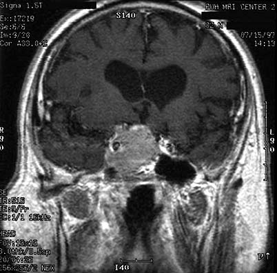

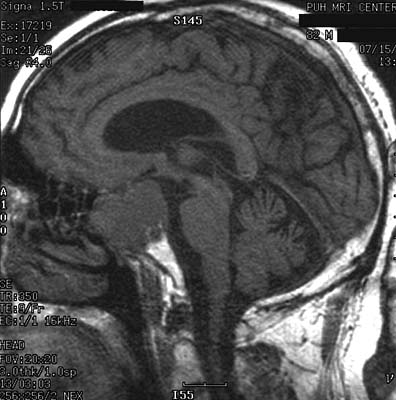
RADIOLOGY AND GROSS DESCRIPTION:
Comparison was made to the prior magnetic resonance (MR) scan of the pituitary gland dated 4-21-97. There was an overall increase in the size of the large pituitary tumor, particularly along its anterior and superior aspect, which measured 4 x 3.5 x 3.5 cm (AP x transverse x long). The optic chiasm could no longer be separately identified from the mass. There was obliteration of the suprasellar cistern, and the pituitary stock was deviated to the left. Also, extension of the tumor to the right cavernous sinus resulted in encasement of the right internal carotid artery. Post operative change with heterogeneous signal material within the sphenoid sinus was seen. Grossly, the surgical resection specimen was received as multiple irregular fragments of pink-tan soft tissue.