
IMAGING STUDIES:
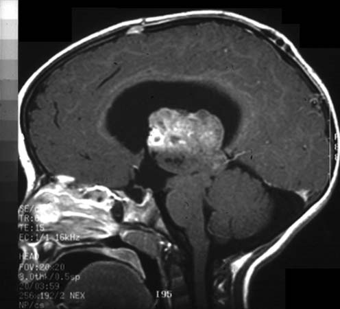
Neuroradiologic images at the time of presentation (Image 01: sagittal T1 weighted image after gadopentetate dimeglumine contrast injection; Image 02: horizontal T2 weighted image). Both pictures show an irregularly shaped mass occupying the third ventricle, with apparent involvement of the septum (Image 02). There is signal heterogeneity with intermediate and high signal and irregular enhancement after contrast injection (Image 01). The tumor compresses the quadrigeminal plate and the lateral ventricles show dilatation with transependymal CSF flow. Fourth ventricle is normal in size.