![]() Contributed by Ronald L. Hamilton, MD.
Contributed by Ronald L. Hamilton, MD.
This nine-year-old previously healthy girl presented with nausea, vomiting and hydrocephalus.
RADIOLOGIC DESCRIPTION:
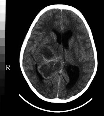

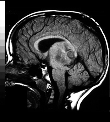
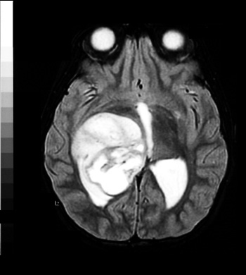
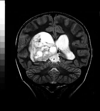
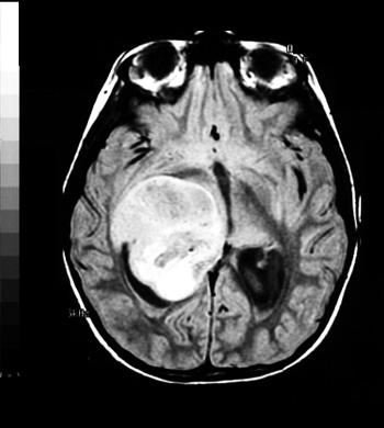
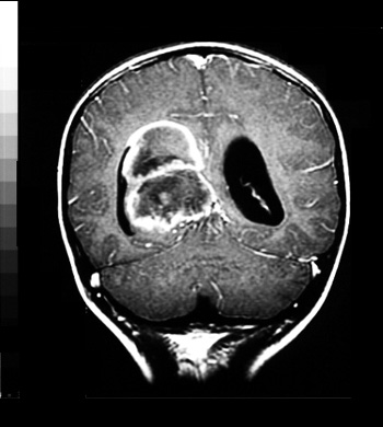
Axial CT scans show a large mass in the right thalamus.CT scan with contrast show some enhancement, especially around the edges of the mass. T-1 weighted sagittal MRI images demonstrate the relationship of the mass to the brainstem and thalamus. Axial T-2 weighted and coronal T-2 weighted illustrate the large size and heterogenous character of the mass, which appears to have cystic or necrotic areas. Proton-density MRI images also demonstrate the variable nature of the thalamic mass. Contrast enhanced MRI coronal images demonstrate the variable contrast enhancement of the mass.
A craniotomy and resection was performed.