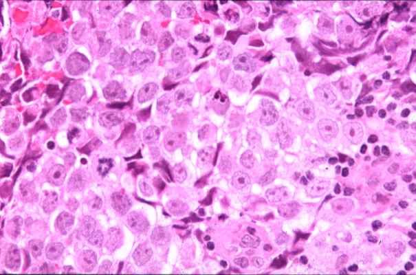
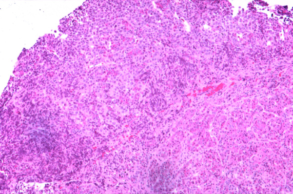
H&E stained sections demonstrate sheets of large, round neoplastic cells separated by fine fibrovascular septa. The tumor cells are characterized by clear to amphophilic cytoplasm and large, round to irregular, creased nuclei containing peripherally distributed chromatin and a single prominent nucleolus. Mitotic figures are common. In some areas there are scattered small cells resembling lymphocytes. In a few places the tumor insinuates into the pituitary parenchyma. There is no necrosis. Fragments of uninvolved pituitary parenchyma appear unremarkable.
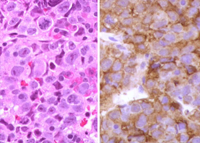
IMMUNOHISTOCHEMISTRY, SPECIAL STAINS AND OTHER STUDIES