Case 938 -- A Post-menopausal Female with Bleeding and Vaginal Bulge

 Contributed by Kavita Varma, MD
CLINICAL HISTORY
Post-menopausal female presented with bleeding and vaginal bulge. On pelvic examination her third degree uterine prolapse was noted. Ultrasound of the pelvis revealed 71 x 44 x 44mm uterus. The endometrial lining measured 4.3mm in thickness. There were two leiomyomas one, Right, posterior, intramural that measured 13 x 14 x 13mm. and second was left, fundal, subserosal that measured 16 x 16 x 14mm.Bilateral ovaries were unremarkable. The patient underwent supracervical hysterectomy and bilateral salpingectomy.
PATHOLOGICAL FINDINGS
- Grossly: The endometrial lining was 0.1 cm thick. The myometrium demonstrated several pale tan well circumscribed whorled nodules ranging from 0.4 x 0.4 x 0.5 cm to 1.6 x 1.2 x 1.1 cm.
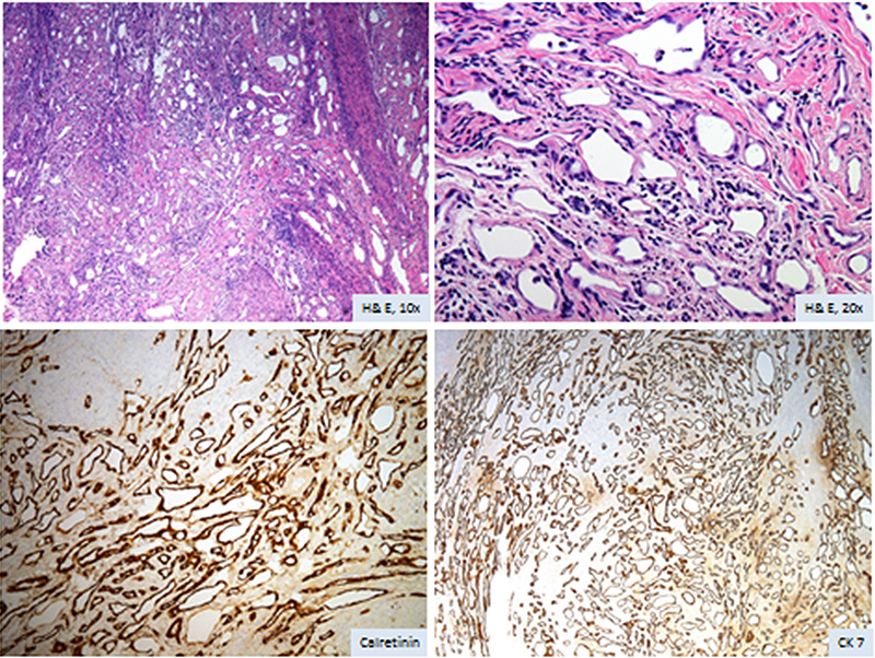
- Microscopically: Sections from one of the nodules revealed adenomatoid pattern with small tubules seen infiltrating into the myometrium. The tubules were lined by cuboidal to flat cells with round nuclei and moderate amount of eosinophilic cytoplasm. No areas of necrosis or increased mitosis were noted. Intervening stroma showed smooth muscle fibres.
- Immunohistochemistry: Tumor cells were strongly positive for calretinin and CK7
FINAL DIAGNOSIS