![]() Contributed by Daniel Marker, MD, PhD and Jennifer Picarsic, MD
Contributed by Daniel Marker, MD, PhD and Jennifer Picarsic, MD
CLINICAL HISTORY
The patient is an adolescent nulligravid female with no significant previous medical history who presented to the emergency department on instructions from her primary care physician for the primary complaint of abdominal fullness and distension. Over the previous four months, she had noticed a "fuller appearance" to her lower abdomen. This change in appearance was not associated with any pain, cramping, or changes in her menstrual cycles, which continued to be regular.
Her initial physical exam in the emergency department revealed a distended, non-tender lower abdomen with a large palpable midline mass. The remainder of her physical exam was unremarkable. Initial laboratory workup was unremarkable, including normal levels AFP, hCG, LDH, CEA-125, and estriol. She underwent ultrasound of the abdomen and CT scan of the chest, abdomen, and pelvis (Figure 1). The CT scan revealed a solid, enhancing, 19 x 12 x 10 cm multilobulated mass arising from the left aspect of the uterus. This mass distorted the endometrial cavity and exerted significant mass effect on the iliac veins, iliac arteries, and distal ureters. The remainder of the abdominal and thoracic CT was unremarkable. The initial interpretation was worrisome for uterine malignancy, such as leiomyosarcoma or other uterine sarcomas. There was also some concern, based on the combined interpretation of the CT and ultrasound, that the mass could be arising from one or both ovaries. After discussion with the patient and her family of the above findings, and because the patient was clinically stable, she was discharged to home with short interval follow-up. A subsequent MRI of the abdomen and pelvis two days later as an outpatient showed normal appearing ovaries and re-demonstrated the large uterine mass (Figure 2).
After extensive interdisciplinary discussion, including consultation with gynecologic-oncologic surgeons, the decision was made to perform an open biopsy for tissue diagnosis, as opposed to resecting the entire mass. The surgery was performed 5 days after presentation. The patient tolerated the procedure well, and the specimen was sent for pathologic diagnosis. The tissue was described grossly as "tan-pink and mildly dense… [with] a smooth membranous delimiting layer."
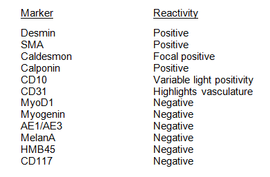
Representative images of the H&E sections and select immunostains are shown, confirming smooth muscle lineage of the tumor (Figures 3, 4, 5, 6, 7, 8, 9, and 10). The tumor shows variable high cellularity with prominent vascularity. The overall cytology is bland, ranging from small ovoid cells to spindle cells with tapered nuclei. No atypical nuclear features are present. The growth pattern is variable, with some areas showing whorls around vasculature and others showing a single file growth pattern in dense collagenous stroma. A single mitosis is identified in 20 high powered fields and the Ki67 proliferation index is less than 1%. No atypical mitoses are identified. No areas of necrosis are present. The full immunophenotype of the tumor is as follows:
