
![]() Contributed by Thomas Pearce, MD, PhD and Juan Xing, MD
Contributed by Thomas Pearce, MD, PhD and Juan Xing, MD
CASE PRESENTATION
A young man in his early 20s presented to an orthopedic oncologist for evaluation of a mass on the lateral aspect of his right lower leg. He gave a history of the mass growing slowly but progressively over the course of approximately one year. The lesion itself was non-tender, but did cause the patient some foot and ankle pain when he stood for long periods of time, and caused intermittent tingling of the leg and foot distally. The patient denied any history of trauma or prior surgical intervention at the site.
On exam, a 2 x 1.5 cm mass was visible approximately 15 cm above the lateral malleolus. The mass was non-tender to palpation, firm, and fixed to the deep tissues. Tinel's sign was positive. The overlying skin was unremarkable. A fine needle aspiration (FNA) of the mass was performed (Figures 1 and 2).
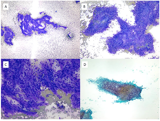
Figure 1: FNA smear preparation. A-B: Tightly cohesive tissue fragments with variable cellularity and dense fibrillary substance (Diff-Quik stain, low power field). C: Tumor cells show wavy nuclei and indistinct cell borders (Diff-Quik stain, high power field). D: PAP stain, low power field.
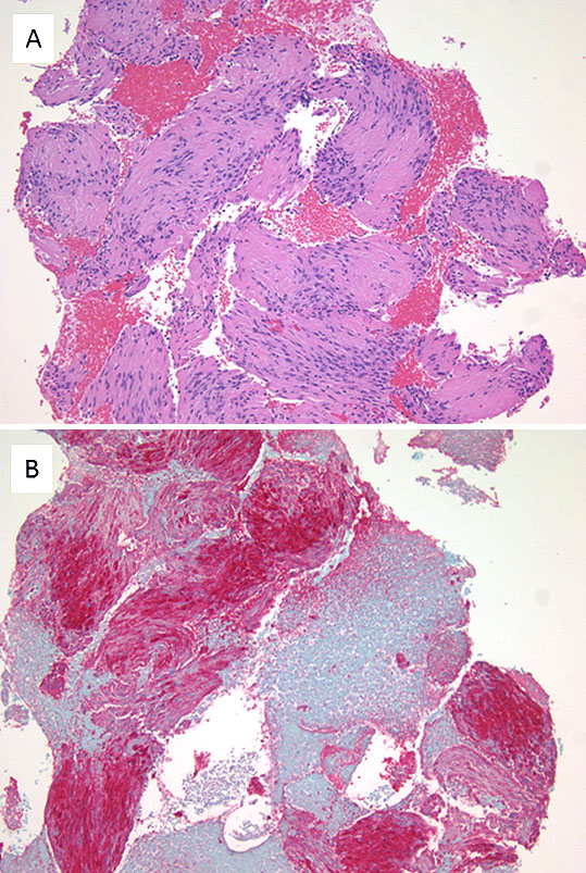
Figure 2: Cell block preparation. A: Palisaded nuclei surrounding dense eosinophilic cytoplasmic processes are appreciated (H&E; low power field). B: The cells stain strongly with S100 by immunohistochemistry (red chromogen).