
![]() Contributed by Lama Farchoukh, MD, Amin Milon, MD and Dr. Anil Parwani, MD, PhD
Contributed by Lama Farchoukh, MD, Amin Milon, MD and Dr. Anil Parwani, MD, PhD
CLINICAL HISTORY
The patient is a woman in her 70s with a remote history of hysterectomy and unilateral salpingo-oophorectomy for "likely benign disease" presented with dull and progressively worsening lower abdominal pain, followed by hematuria.
IMAGING STUDIES
)
)
CT abdomen with contrast showed distended urinary bladder measuring 15.0 x 12.1cm (Figure 1) and severe diffuse bladder wall thickening with bladder trigone inseparable from the anterior wall of the thickened vagina concerning for invasion (Figure 2). Cystoscopic examination showed severe hemorrhagic cystitis and necrotic tissue. The patient underwent a transurethral resection of this mass.
TRANSURETHRAL RESECTION OF URINARY BLADDER LESION
)
)
Histologic sections of the transurethral resection specimen demonstrated submucosal infiltration by architecturally complex glands with enlarged, raisinoid and hyperchromatic nuclei as well as prominent nucleoli in comparison to the surrounding urothelium. A subset of these cells had clear cytoplasm (Figures 3A and 3B). Paraffin section immunohistochemical analysis demonstrated that the abnormal cell population was strongly and diffusely immunoreactive for PAX-8 (Figure 4A), hepatocyte nuclear factor beta-1 (HNF-B1) (Figure 4B), cytokeratin 7 (CK7) (Figure 4C) and alpha-methylacyl CoA racemase (P504S) (Figure 4D), and showed membranous immunoreactivity with high molecular weight cytokeratin (HMWK) (Figure 4E) and CAM 5.2 with weaker intensity than that expressed by adjacent urothelium. The tumor cells were negative for CK20, cdx-2, p63, TTF-1, WT-1, ER, PR, GCDFP-15 and mammaglobin. Metastatic workup, including CT scan, bone scan and liver function tests (LFTs) showed no obvious evidence of metastatic disease. The patient underwent an anterior exenteration.
GROSS DESCRIPTION
)
Anterior exenteration with excision of the remaining ovary and fallopian tube demonstrated a 6.7 cm papillary mass in the inferior aspect of the posterior bladder wall and proximal urethra (Figure 5A), extending through the detrusor muscle into the deep anterior vaginal wall (Figure 5B).
HISTOLOGICAL FINDINGS
)
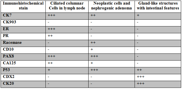
Histologic sections showed features similar to those from the patient's prior transurethral tumor resection; however, a number of adjacent elements were identified, including in-situ adenocarcinoma (Figure 6A) and nephrogenic adenoma (Figure 6B). In addition, the malignant glands appeared to transition into a focus of background glands with goblet cells (Figure 6C), and seven out of twenty-one (7/21) lymph nodes were involved by haphazardly arranged and ciliated glandular structures without surrounding endometriotic stroma or hemorrhage (Figure 6D). To further characterize this malignancy, additional immunohistochemical stains were performed on sections with each of the aforementioned elements (Table 1).
IMMUNOHISTOCHEMICAL FINDINGS
)
)
)
The malignant cells showed strong expression of p53 and focal immunoreactivity with CA125 and CD10. Both the malignant cell population and the nephrogenic adenoma demonstrated an identical immunophenotype, with immunoreactivity for CK7 (Figure 7A) and P504S (Figure 7B) but no expression of ER (Figure 7C), CK20 (Figure 7D) or cdx-2 (Figure 7E). The background glands were immunoreactive with CK20 (Figure 8A) and cdx-2 (Figure 8B), but not PAX-8 (Figure 8C). The ciliated glandular structures within the lymph nodes were immunoreactive for ER (Figure 9A), PR (Figure 9B) and WT-1 (Figure 9C), but not cdx-2 (Figure 9D) or P504S.