![]() Contributed by: Dorian Infantino, MD
Contributed by: Dorian Infantino, MD
PATIENT HISTORY
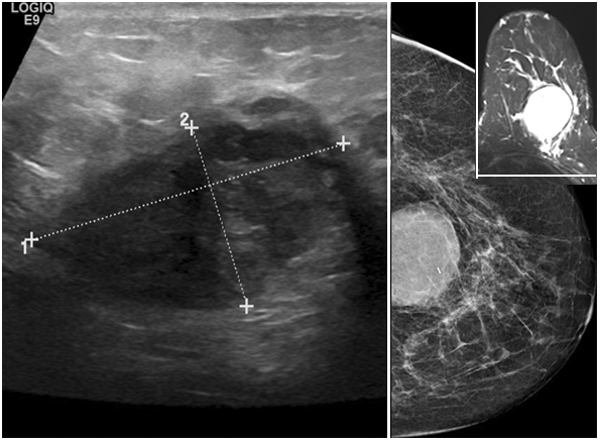
68 year old woman presented with a swollen and tender mass in her left breast. The patient has a history of fibrocystic changes with cyst formation. Diagnostic mammogram and ultrasound showed a 4.8 x 4.8 x 2.7 cm solid and cystic mass with irregular borders and mixed echogenicity located against the chest wall at the 6:00 position approximately 6 cm from the nipple (see below). Core needle biopsy was performed, demonstrating the histopathology shown below, and ER/PR/HER2 immunohistochemistry demonstrated a triple negative phenotype. The patient was evaluated for neoadjuvant therapy and none was performed after proper pathologic diagnosis. The patient then underwent left breast lumpectomy with sentinel lymph node biopsy (see below).
IMAGING
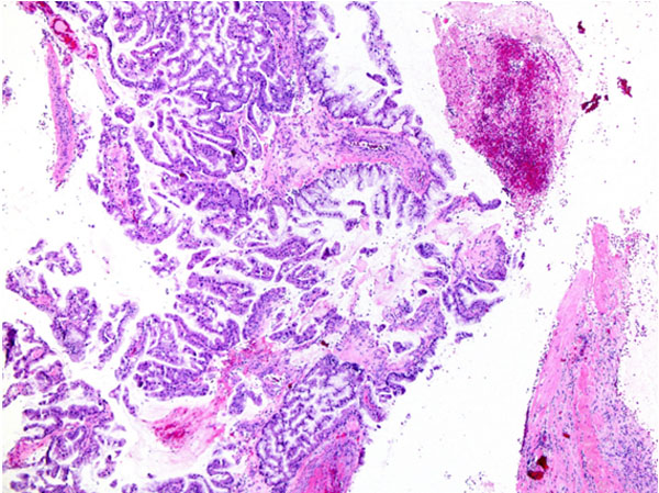
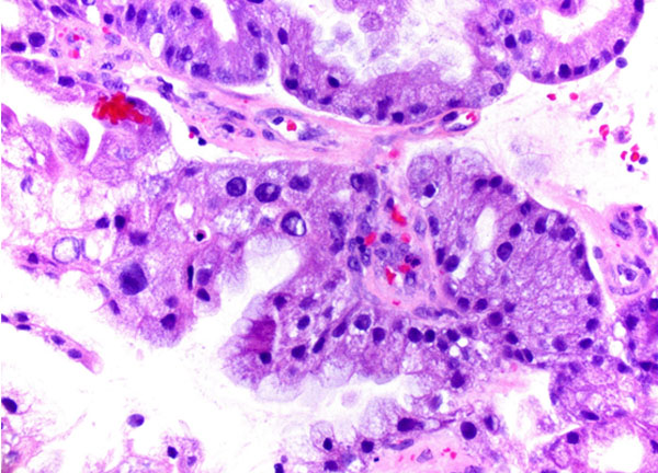
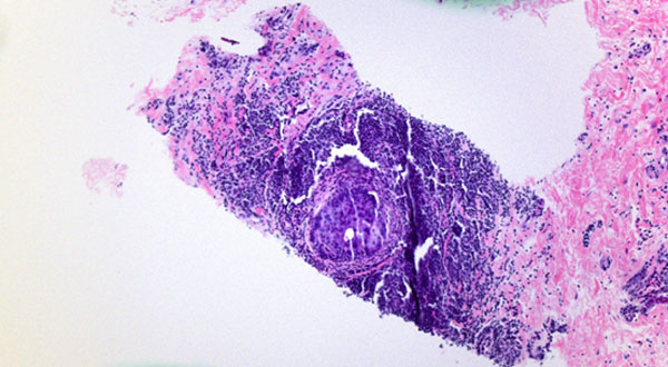
HISTOLOGY
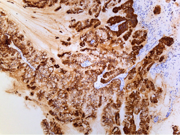
Core Biopsy
Cystically dilated spaces lined by papillary, tufted epithelium with basally oriented nuclei and intracytoplasmic mucin.
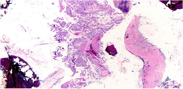
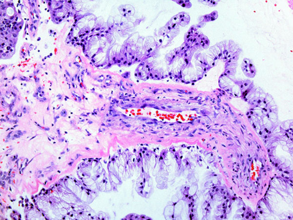
Variable nuclear atypia (from bland nuclei to moderate atypia) and cytoplasmic eosinophilia.
Focal ductal carcinoma in situ with periductal chronic inflammation.
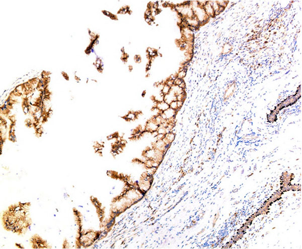
IMMUNOHISTOCHEMISTRY
CK7: Positive
Mammoglobin: Positive
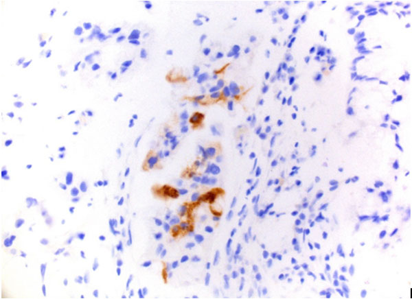
Negative stains: CK20, CDX2, PAX8, WT1, GCDFP15, GATA3, ER, PR, HER2
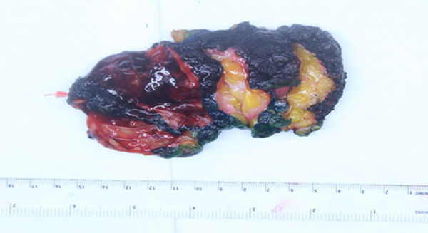
EXCISION
Left Breast Lumpectomy
Gross: 6.2 x 4.4 x 4.0 cm cystic lesion, filled with opaque brown viscous fluid.
Intracellular and extracellular mucin
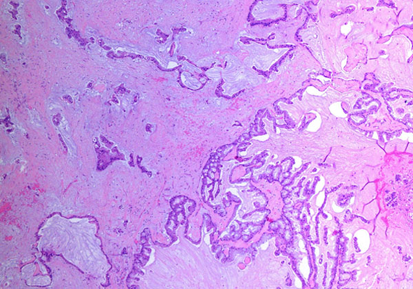
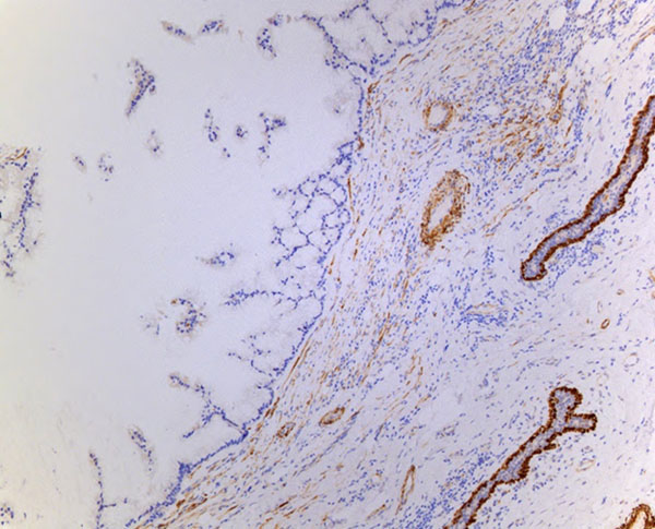
Myoepithelial markers negative: P63 and calponin