
![]() Contributed by Virginia Miller, MD and Jon M. Davison, MD, Department of Pathology, University of Pittsburgh Medical Center
Contributed by Virginia Miller, MD and Jon M. Davison, MD, Department of Pathology, University of Pittsburgh Medical Center
CLINICAL HISTORY
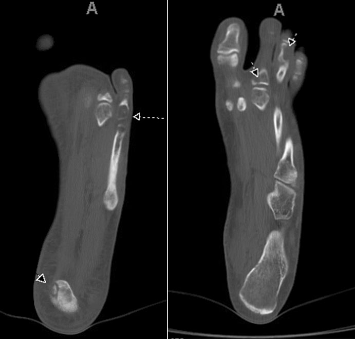
A Caucasian male in his early 20's presented with a cutaneous, soft tissue mass of the right lower extremity as well as vascular abnormalities of the mid-3rd phalange. Subsequent pathologic fracture of the proximal 2nd phalange, led to the discovery of multiple osseous lesions of the left foot (below). Excision of the lesions showed similar histologic features in both the soft tissue and bone sites. Long-term follow up was characterized by multiple bone (tibia) and soft tissue recurrences within the tissues of the right lower limb.
IMAGING
HISTOLOGY

Periphery of cutaneous lesion with infiltrative border
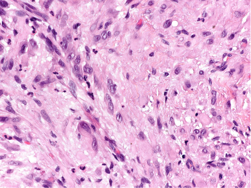
High-power cutaneous lesion with cytologic/nuclear detail

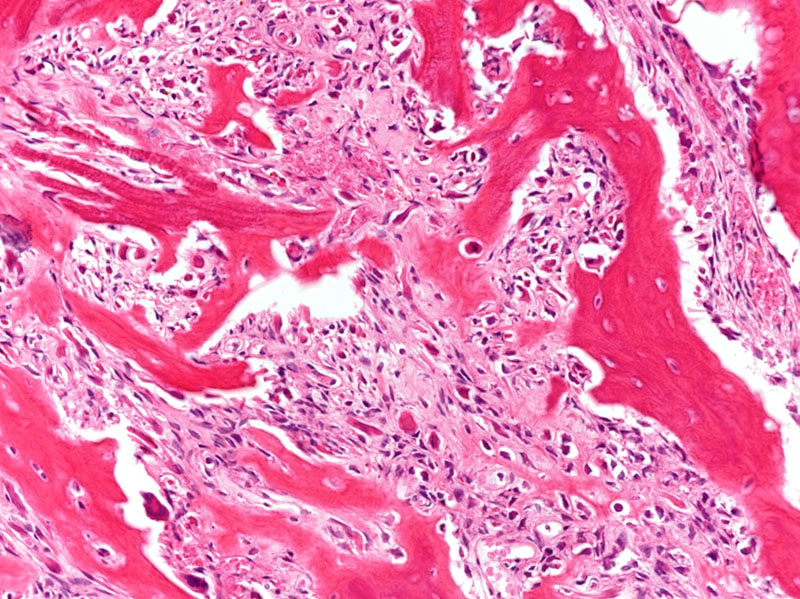
High-power bony lesion, post decal (2nd phalange)
STAINS
MOLECULAR FINDINGS
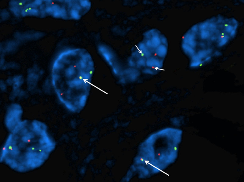
FISH
FOSB-1/SERPINE-1 t(7;19) gene fusion.