![]() Contributed by Laura Favazza, DO and Arivarasan Karunamurthy, MD, Department of Pathology, University of Pittsburgh Medical Center
Contributed by Laura Favazza, DO and Arivarasan Karunamurthy, MD, Department of Pathology, University of Pittsburgh Medical Center
CLINICAL HISTORY
This is a female in her late 40s with a history of uveal melanoma in her left eye status post plaque radiation, four years ago. She now presents with multiple subcutaneous lesions, as well as lung and liver lesions. A subcutaneous lesion on the back was biopsied.
SURGICAL PATHOLOGY REPORT
SOFT TISSUE, RIGHT BACK, EXCISION: METASTATIC MELANOMA, CONSISTENT WITH A UVEAL PRIMARY, INVOLVING FIBROADIPOSE TISSUE (see comment).
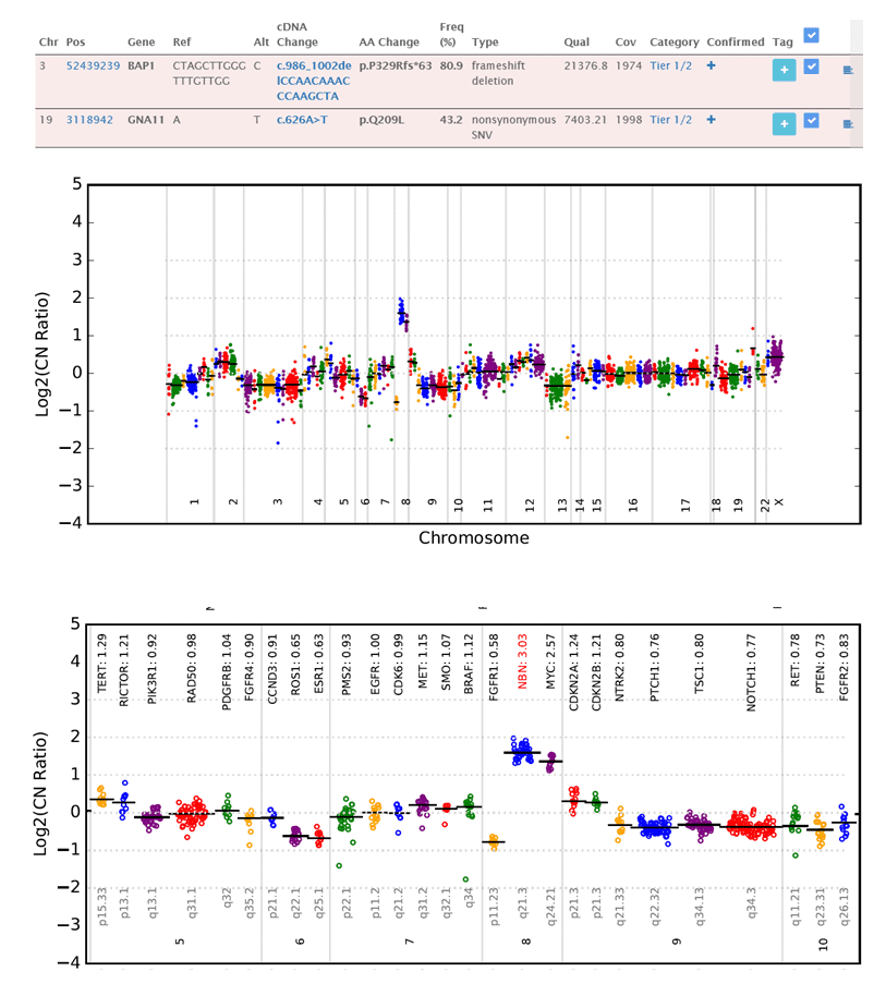
COMMENT: Per the patient's electronic medical record, a history of a widely metastatic uveal melanoma is noted. The biopsy specimen consists of a pigmented spindle cell neoplasm. An immunohistochemical stain for Sox10 is positive within the neoplastic cells; while, BAP1 shows loss of nuclear expression. Based on the histologic findings and immunoprofile, the neoplasm is consistent with a metastatic melanoma from the patient's known uveal primary.
Oncomine testing is performed.